While Microsoft has patched its software in recent days, cybersecurity researchers have already detected breaches on more than 100 servers representing 60 victims thus far
Get the latest international news and world events from around the world.
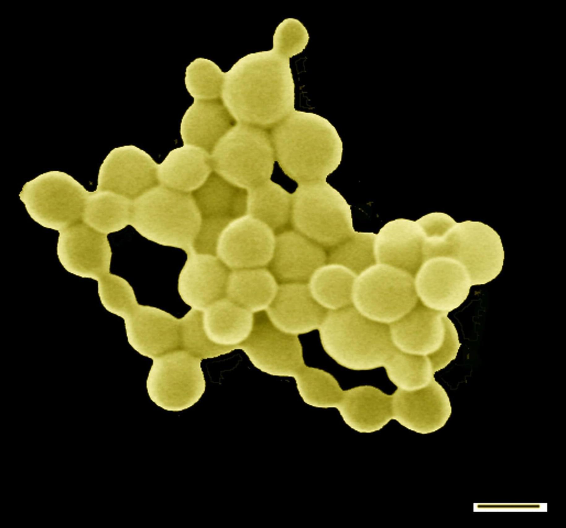
Researchers Reveal How Bacteria Can Produce Gold
High concentrations of heavy metals, like copper and gold, are toxic for most living creatures. This is not the case for the bacterium C. metallidurans, which has found a way to extract valuable trace elements from a compound of heavy metals without poisoning itself. One interesting side-effect: the formation of tiny gold nuggets. A team of researchers from Martin Luther University Halle-Wittenberg (MLU), the Technical University of Munich (TUM) and the University of Adelaide in Australia has discovered the molecular processes that take place inside the bacteria. The group presented their findings in the renowned journal Metallomics published by the Royal Society of Chemistry.
A team of researchers reveal how bacterium C. metallidurans extracts valuable trace elements from a compound of heavy metals without poisoning itself, and thereby produces gold particles.
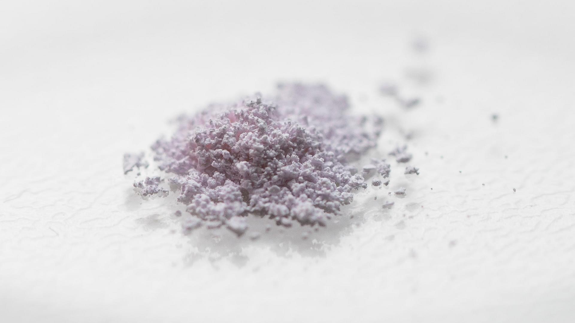
Breakthrough method purifies rare earths element with just water
A rare earth breakthrough could rewrite the rules of recycling.
Scientists at IOCB Prague have developed a cleaner, smarter way to recover these critical elements, crucial to technologies from smartphones to wind turbines.
The technique can efficiently extract metals like neodymium and dysprosium from discarded magnets, bypassing the toxic solvents and waste generated by conventional processes.
With global demand for rare earths soaring, the need for sustainable recovery methods has never been greater.
ICOB Prague’s chelator tech separates rare earths cleanly—and just revealed holmium’s surprise EV comeback.
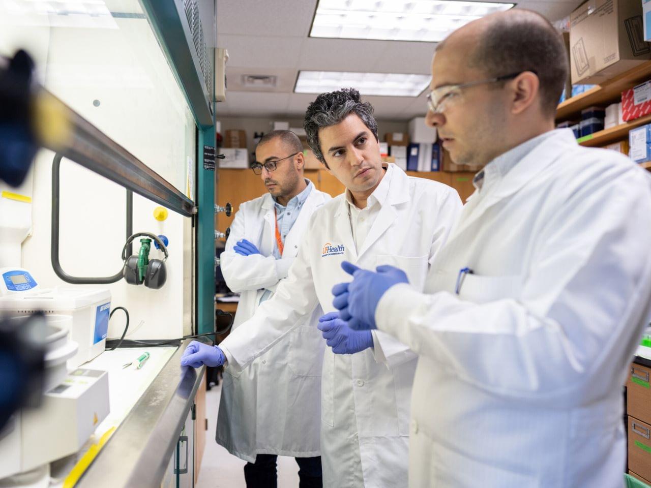
Surprising finding could pave way for universal cancer vaccine
An experimental mRNA vaccine boosted the tumor-fighting effects of immunotherapy in a mouse-model study, bringing researchers one step closer to their goal of developing a universal vaccine to “wake up” the immune system against cancer.
Published today in Nature Biomedical Engineering, the University of Florida study showed that like a one-two punch, pairing the test vaccine with common anticancer drugs called immune checkpoint inhibitors triggered a strong antitumor response.
A surprising element, researchers said, was that they achieved the promising results not by attacking a specific target protein expressed in the tumor, but by simply revving up the immune system — spurring it to respond as if fighting a virus. They did this by stimulating the expression of a protein called PD-L1 inside of tumors, making them more receptive to treatment. The research was supported by multiple federal agencies and foundations, including the National Institutes of Health.
Sc: What research can be furthered?
What has not yet been tried? These are the questions that Inserm research director Nicolas L’Heureux has asked himself every day for a long time, « like a game ». Which means that from very early on he had the idea of pushing the limits of vascular tissue engineering – a field in which he had begun working when doing his M.Sc. « When performing a cardiac or other type of bypass, preference is given to using the patient’s own vessels that are taken from one place and transplanted into another, more critical, one. An autologous graft continues to remain the best solution, but it is a limited resource. » Diseases such as stroke, hyperlipidemia, and thrombosis, which have the particularity of being systemic – in which they attack all vessels to varying degrees –, as well as aging, weaken our vessels. And the earlier the need for surgery, the greater the likelihood of a second intervention. « A transplanted artery will withstand an average of ten years and a vein six to seven years. » Which just leaves synthetic grafts. https://www.inserm.fr/en/news/nicolas-lheureux-artificial-bl…iological/
See relevant content for m.
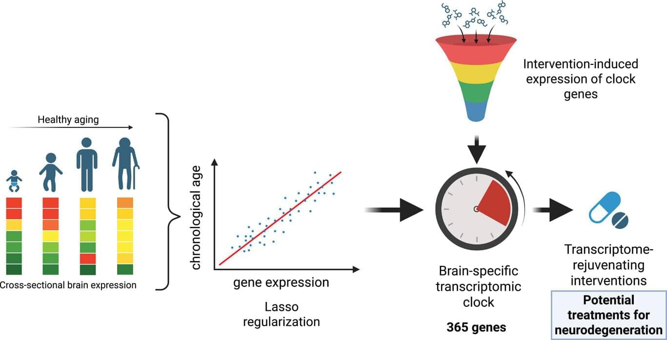
Computational clock identifies compounds that may rejuvenate aging brain cells
What if there was a way to make aging brain cells younger again? An international research team from Spain and Luxembourg recently set out to address this question. After developing an aging clock capable of assessing the biological age of the brain, they used it to identify possible brain-rejuvenating interventions. The computational tool they created, recently presented in the journal Advanced Science, constitutes a valuable resource to find compounds with therapeutic potential for neurodegenerative diseases.
As the world population is aging rapidly, with over two billion people projected to be above the age of 60 by 2050, age-related brain disorders are on the rise. Living longer but in poor health is not only a daunting prospect, it also places a substantial burden on health care systems worldwide. The idea of being able to counteract the functional decline of our brain through rejuvenating interventions therefore sounds promising.
The question is, how can we identify compounds that have the potential to efficiently rejuvenate brain cells and to protect the aging population from neurodegeneration? Prof. Antonio Del Sol and his teams of computational biologists, based both at CIC bioGUNE, member of BRTA, and the Luxembourg Centre for Systems Biomedicine (LCSB) from the University of Luxembourg, used their machine learning expertise to tackle the challenge.
Scientists challenge theory that human eye evolution was driven by cooperation
Humans are often said to be the only primates with “whites of the eyes,” evolved for social communication. But a new study challenges that idea, arguing that the theory lacks evidence and oversimplifies the diversity of primate eye pigmentation.

Trestle Bio Announces Research Collaboration with Humacyte
Researchers have created a cement-based material that does more than just provide structural support—it can generate and store electricity. This breakthrough could mark a turning point for future infrastructure in smart cities.
The material is a cement-hydrogel composite developed by a team led by Professor Zhou Yang at Southeast University in China. The team took inspiration from the layered structure inside plant stems to create a material that can harness thermal energy and convert it into electricity.
This is a repost. I think Andrew posted it earlier.
Researchers developed a cement-hydrogel composite that can generate and store power, paving the way for self-powered smart infrastructure.