Toggle light / dark theme

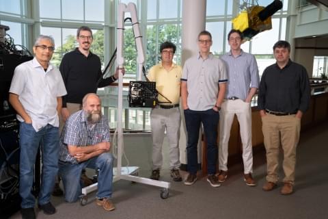
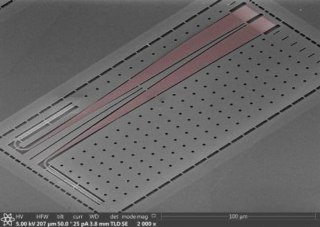
Researchers from the University of Tsukuba have shown how adding a tiny resonator structure to an ultrafast electron pulse detector reduced the intensity of terahertz radiation required to characterize the pulse duration (ACS Photonics, “Streaking of a Picosecond Electron Pulse with a Weak Terahertz Pulse”).

To study proteins—for example, when determining the mechanisms of their biological actions—researchers need to understand the motion of individual atoms within a sample. This is difficult not just because atoms are so tiny, but also because such rearrangements usually occur in picoseconds—that is, trillionths of a second.

One method to examine these systems is to excite them with an ultrafast blast of laser light, and then immediately probe them with a very short electron pulse. Based on the way the electrons scatter off the sample as a function of the delay time between the laser and electron pulses, researchers can obtain a great deal of information about the atomic dynamics. However, characterizing the initial electron pulse is difficult and requires complex setups or high-powered THz radiation.

What are #dopamine, #serotonin, norepinephrine, glutamate, #GABA, acetylcholine? What does dopamine do?
📢SUBSCRIBE and click the BELL to get notified when new videos are uploaded.
💲 AllCEUs.com Unlimited continuing education CEUs $59
💻 Online course based on this video can be found at AllCEUs.com/NEURO-CEU https://www.allceus.com/member/cart/index/product/id/96/c/
⭐ Specialty Certificate Programs for Case Management and Counselor Certification beginning at $89 https://AllCEUs.com/certificate-tracks.

Join this channel to get access to perks:
https://www.youtube.com/channel/UCAE3JJi8tX7gfhZEXCUGd_A/join.

NOTE: ALL VIDEOS are for educational purposes only and are NOT a replacement for medical advice or counseling from a licensed professional.

Video by Dr. Dawn Elise Snipes on integrative behavioral health approaches including counseling techniques and skills for improving mental health and reducing mental illness.

Human-like articulated neural avatars have several uses in telepresence, animation, and visual content production. These neural avatars must be simple to create, simple to animate in new stances and views, capable of rendering in photorealistic picture quality, and simple to relight in novel situations if they are to be widely adopted. Existing techniques frequently use monocular films to teach these neural avatars. While the method permits movement and photorealistic image quality, the synthesized images are constantly constrained by the training video’s lighting conditions. Other studies specifically address the relighting of human avatars. However, they do not provide the user control over the body stance. Additionally, these methods frequently need multiview photos captured in a Light Stage for training, which is only permitted in controlled environments.

Some contemporary techniques seek to relight dynamic human beings in RGB movies. However, they lack control over body posture. They need a brief monocular video clip of the person in their natural location, attire, and body stance to produce an avatar. Only the target novel’s body stance and illumination information are needed for inference. It is difficult to learn relightable neural avatars of active individuals from monocular RGB films captured in unfamiliar surroundings. Here, they introduce the Relightable Articulated Neural Avatar (RANA) technique, which enables photorealistic human animation in any new body posture, perspective, and lighting situation. It first needs to simulate the intricate articulations and geometry of the human body.

The texture, geometry, and illumination information must be separated to enable relighting in new contexts, which is a difficult challenge to tackle from RGB footage. To overcome these difficulties, they first use a statistical human shape model called SMPL+D to extract canonical, coarse geometry, and texture data from the training frames. Then, they suggest a unique convolutional neural network trained on artificial data to exclude the shading information from the coarse texture. They add learnable latent characteristics to the coarse geometry and texture and send them to their proposed neural avatar architecture, which uses two convolutional networks to produce fine normal and albedo maps of the person underneath the goal body posture.

Leading Canada’s Bio-Safety & Security R&D — Dr. Loren Matheson PhD, Defence Research and Development Canada, Department of National Defence.


Dr. Loren Matheson, Ph.D. is a Portfolio Manager at the Center For Security Science, at Defence Research and Development Canada (DRDC — https://www.canada.ca/en/defence-research-development.html), which is a special operating agency of the Department of National Defence, whose purpose is to provide the Canadian Armed Forces, other government departments, and public safety and national security communities with knowledge and technology.

With a focus on the chemical and biological sciences at DRDC, Dr. Matheson develops and leads safety and security R&D projects with government partners, industry and academia. In addition, she spearheaded an effort to establish a virtual symposium series, developed communications products to explain their program to national and international partners, and helped established a science communication position.

New video: Dr. Aaron Cravens, CEO of Revel Pharmaceuticals at #RejuvenationStartupSummit 2022 about developing designer enzymes to treat diseases of aging, e.g. glucosepane crosslink breakers to prevent hardening of tissues such as muscle, skin, and arteries leading to increased blood pressure, vascular damage, and wrinkling of the skin.

Enjoy →

For more summit videos and regular updates, visit: youtube.com/user/foreverhealthyorg


In addition to laser-assisted bioprinting, other light-based 3D bioprinting techniques include digital light processing (DLP) and two-photon polymerization (TPP)-based 3D bioprinting. DLP uses a digital micro-mirror device to project a patterned mask of ultraviolet (UV)/visible range light onto a polymer solution, which in turn results in photopolymerization of the polymer in contact [56, 57]. DLP can achieve high resolution with rapid printing speed regardless of the layer’s complexity and area. In this method of 3D bioprinting, the dynamics of the polymerization can be regulated by modulating the power of the light source, the printing rate, and the type and concentrations of the photoinitiators used. TPP, on the other hand, utilizes a focused near-infrared femtosecond laser of wavelength 800 nm to induce polymerization of the monomer solution [56]. TPP can provide a very high resolution beyond the light diffraction limit since two-photon absorption only happens in the center region of the laser focal spot where the energy is above the threshold to trigger two-photon absorption [56].

The recent development of the integrated tissue and organ printer (ITOP) by our group allows for bioprinting of human scale tissues of any shape [45]. The ITOP facilitates bioprinting with very high precision; it has a resolution of 50 μm for cells and 2 μm for scaffolding materials. This enables recapitulation of heterocellular tissue biology and allows for fabrication of functional tissues. The ITOP is configured to deliver the bioink within a stronger water-soluble gel, Pluronic F-127, that helps the printed cells to maintain their shape during the printing process. Thereafter, the Pluronic F-127 scaffolding is simply washed away from the bioprinted tissue. To ensure adequate oxygen diffusion into the bioprinted tissue, microchannels are created with the biodegradable polymer, polycaprolactone (PCL). Stable human-scale ear cartilage, bone, and skeletal muscle structures were printed with the ITOP, which when implanted in animal models, matured into functional tissue and developed a network of blood vessels and nerves [45]. In addition to the use of materials such as Pluronic F-127 and PCL for support scaffolds, other strategies for improving structural integrity of the 3D bioprinted constructs include the use of suitable thickening agents such as hydroxyapatite particles, nanocellulose, and Xanthan and gellan gum. Further, the use of hydrogel mixtures instead of a single hydrogel is a helpful strategy. For example, the use of gelatin-methacrylamide (GelMA)/hyaluronic acid (HA) mixture instead of GelMA alone shows enhanced printability since HA improves the viscosity of mixture while crosslinking of GelMA retains post-printing structural integrity [58].

To date, several studies have investigated skin bioprinting as a novel approach to reconstruct functional skin tissue [44, 59,60,61,62,63,64,65,66,67]. Some of the advantages of fabrication of skin constructs using bioprinting compared to other conventional tissue engineering strategies are the automation and standardization for clinical application and precision in deposition of cells. Although conventional tissue engineering strategies (i.e., culturing cells on a scaffold and maturation in a bioreactor) might currently achieve similar results to bioprinting, there are still many aspects that require improvements in the production process of the skin, including the long production times to obtain large surfaces required to cover the entire burn wounds [67]. There are two different approaches to skin bioprinting: in situ bioprinting and in vitro bioprinting. Both these approaches are similar except for the site of printing and tissue maturation. In situ bioprinting involves direct printing of pre-cultured cells onto the site of injury for wound closure allowing for skin maturation at the wound site. The use of in situ bioprinting for burn wound reconstruction provides several advantages, including precise deposition of cells on the wound, elimination of the need for expensive and time-consuming in vitro differentiation, and the need for multiple surgeries [68]. In the case of in vitro bioprinting, printing is done in vitro and the bioprinted skin is allowed to mature in a bioreactor, after which it is transplanted to the wound site. Our group is working on developing approaches for in situ bioprinting [69]. An inkjet-based bioprinting system was developed to print primary human keratinocytes and fibroblasts on dorsal full-thickness (3 cm × 2.5 cm) wounds in athymic nude mice. First, fibroblasts (1.0 × 10⁵ cells/cm2) incorporated into fibrinogen/collagen hydrogels were printed on the wounds, followed by a layer of keratinocytes (1.0 × 10⁷ cells/cm2) above the fibroblast layer [69]. Complete re-epithelialization was achieved in these relatively large wounds after 8 weeks. This bioprinting system involves the use of a novel cartridge-based delivery system for deposition of cells at the site of injury. A laser scanner scans the wound and creates a map of the missing skin, and fibroblasts and keratinocytes are printed directly on to this area. These cells then form the dermis and epidermis, respectively. This was further validated in a pig wound model, wherein larger wounds (10 cm × 10 cm) were treated by printing a layer of fibroblasts followed by keratinocytes (10 million cells each) [69]. Wound healing and complete re-epithelialization were observed by 8 weeks. This pivotal work shows the potential of using in situ bioprinting approaches for wound healing and skin regeneration. Clinical studies are currently in progress with this in situ bioprinting system. In another study, amniotic fluid-derived stem cells (AFSCs) were bioprinted directly onto full-thickness dorsal skin wounds (2 cm × 2 cm) of nu/nu mice using a pressure-driven, computer-controlled bioprinting device [44]. AFSCs and bone marrow-derived mesenchymal stem cells were suspended in fibrin-collagen gel, mixed with thrombin solution (a crosslinking agent), and then printed onto the wound site. Two layers of fibrin-collagen gel and thrombin were printed on the wounds. Bioprinting enabled effective wound closure and re-epithelialization likely through a growth factor-mediated mechanism by the stem cells. These studies indicate the potential of using in situ bioprinting for treatment of large wounds and burns.

In a first-of-its-kind clinical trial, a human has received a 3D-bioprinted ear implant grown from the patient’s own living cells – thanks to a technology platform developed by a Cornellian-founded startup company.

This bioengineered breakthrough has the potential to significantly improve the lives of the approximately 1,500 children who are born annually in the U.S. with microtia, a congenital ear deformity. The approach could eventually lead to tissue implants for treating other conditions and traumatic injuries, reconstructive and regenerative therapy, and possibly even the biomanufacture of whole organs.

The company, 3DBio Therapeutics, was founded in 2014 by Dan Cohen ‘04, M.S. ‘07, Ph.D. ‘10, along with Lawrence Bonassar, the Daljit S. and Elaine Sarkaria Professor in Biomedical Engineering and in Mechanical and Aerospace Engineering in the College of Engineering, and Hod Lipson, who taught at Cornell for 14 years and is now a professor at Columbia University.

Honda is pulling away from a design practice that’s (literally) shaped auto making since the ’30s.

The $43 billion company still depends on life-size clay models to evaluate its designs, a tried and true method pioneered by GM designer Harley Earl. But Honda is gradually relying less on the practice, ever since the Coronavirus tore across the globe and resulting lockdowns divided its teams in Los Angeles, Ohio and Japan. The way Honda tells it, those early 2020 travel rules “threatened” its designers’ ability to work with engineers on the ’24 Prologue, creating a window for a deeper dive into virtual reality.