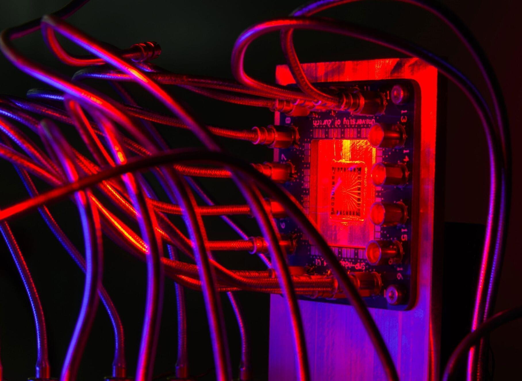
In a step toward making ultra-bright X-ray sources more widely available, an international collaboration led by the University of Michigan—with experiments at the U.K.’s Central Laser Facility—has mapped key aspects of electron pulses that can go on to generate laser-like X-ray pulses.
These X-ray pulses have the potential to advance chemistry, biology, material science and physics by enabling researchers to measure the way molecules behave in great detail. The technique may also be useful in clinical medicine for imaging soft tissues and organs.
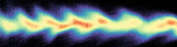
Because the pulses are so short, quadrillionths of a second (femtoseconds) long, they can take snapshots of chemical reactions, revealing the choreography of atoms and molecules, including larger biomolecules such as proteins. These studies are valuable for both basic research, down to quantum mechanics, and applications of chemistry such as drug discovery.


