‘Mini-brains’ grown in the lab allow scientists to study brain wiring without interfering with brains in living people, and now a new study has used these scaled-down models to identify neural signatures associated with schizophrenia and bipolar disorder.
We are providing an unedited version of this manuscript to give early access to its findings. Before final publication, the manuscript will undergo further editing. Please note there may be errors present which affect the content, and all legal disclaimers apply.
Automation and robotics, particularly with the integration of AI, are transforming industries and poised to significantly impact the workforce, but are likely to lead to a reduction in work hours and increased productivity rather than total job destruction.
## Questions to inspire discussion.
Investment & Market Opportunity.
🤖 Q: What is the revenue potential for robotics by 2025? A: ARK Invest projects a $26 trillion global revenue opportunity across household and manufacturing robotics by 2025, driven by convergence of humanoid robots, AI, and computer vision technologies.
💰 Q: How should companies evaluate robot ROI for deployment? A: Robots are worth paying for based on task-specific capabilities delivering 2–10% productivity gains, unlike autonomous vehicles requiring full job performance—Roomba succeeded despite early limitations by being novel and time-saving for specific tasks.
Since the interstellar object (ISO) 3I/ATLAS was first discovered on July 1, 2025, it has garnered much attention, including speculation, hopes and fears that it may somehow contain evidence of technologically advanced civilizations outside of our solar system.
Now, a new paper published on the arXiv preprint server details the findings from radio observations made at the 100-meter Green Bank Telescope as a part of the Breakthrough Listen program, designed to look for signs of alien life. The data were taken on December 18, 2025—the day before the object’s closest approach to Earth, and those hoping for evidence of advanced alien civilizations may not like the results.
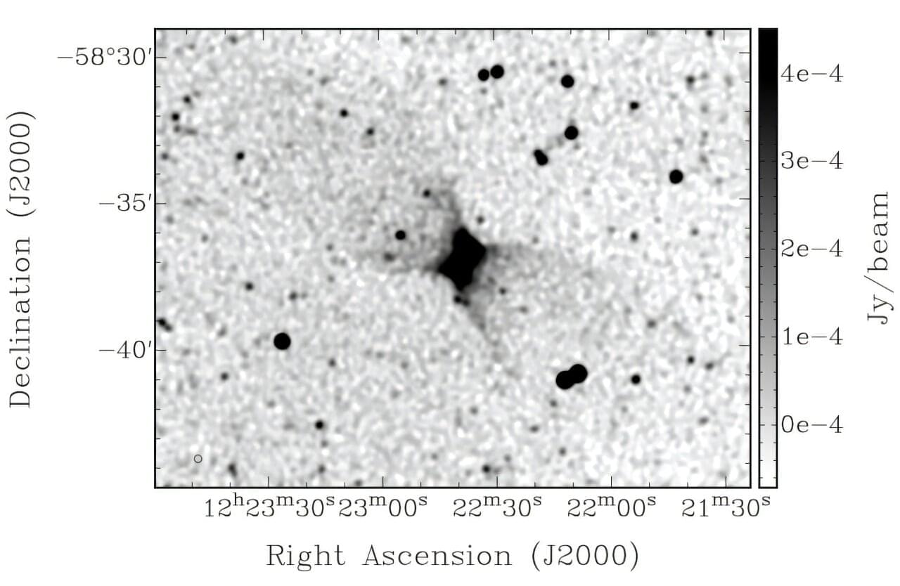
Using the Australian Square Kilometer Array Pathfinder (ASKAP), an international team of astronomers has discovered a spectacular bipolar outflow from the disk of a nearby galaxy known as ESO 130-G012. The finding was reported in a paper published December 17 on the pre-print server arXiv.
ESO 130-G012 is an edge-on galaxy at a distance of some 55 million light years, with an estimated stellar mass of about 11 billion solar masses. The galaxy has a star-formation rate at a level of 0.2 solar masses per year and hosts a black hole approximately 50 million times more massive than the sun.
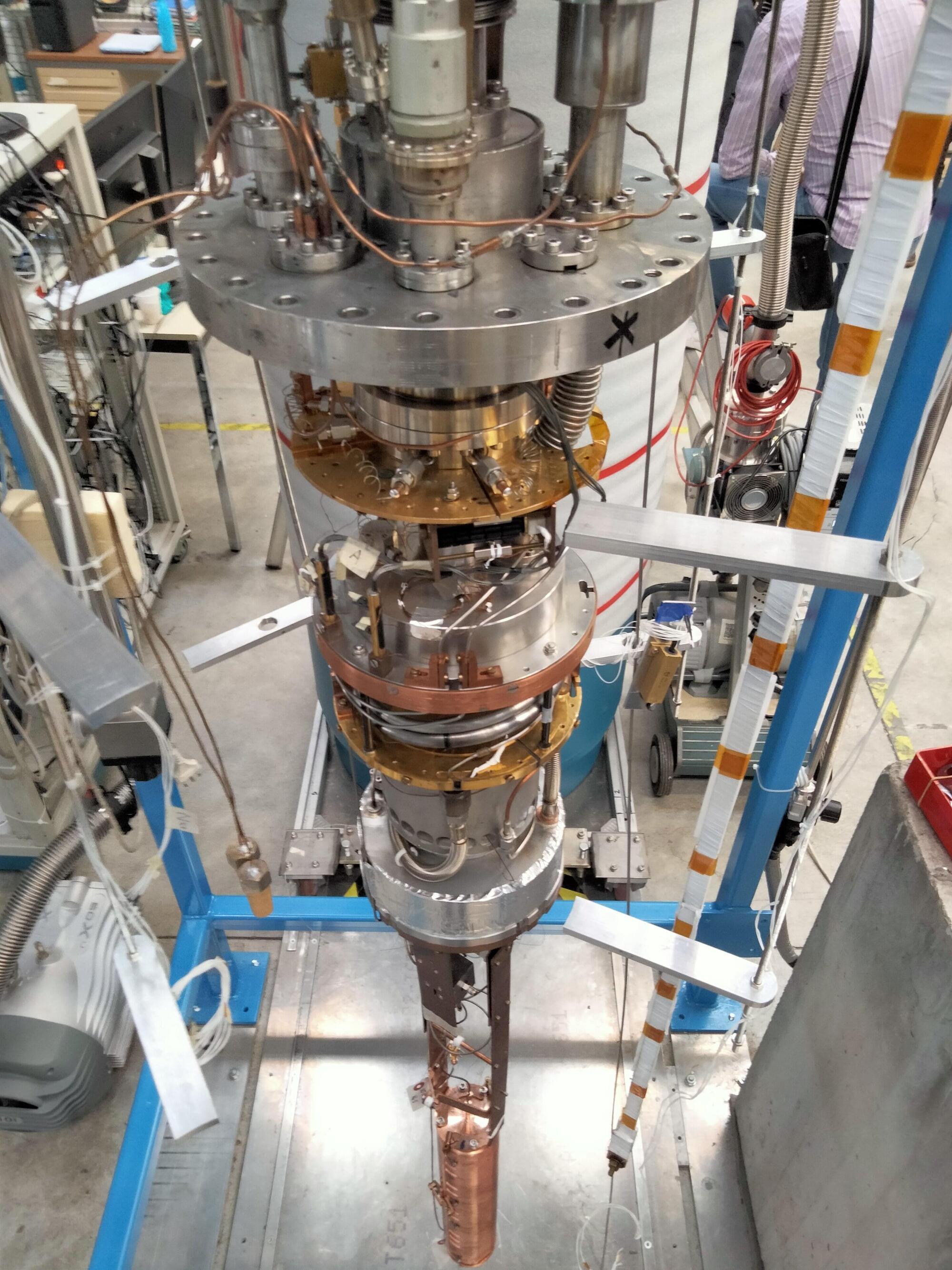
Axions are hypothetical light particles that could solve two different physics problems, as they could explain why some nuclear interactions don’t violate time symmetry and are also promising dark matter candidates. Dark matter is a type of matter that does not emit, reflect or absorb light, and has never been directly observed before.
Axions are very light particles theorized to have been produced in the early universe but that would still be present today. These particles are expected to interact very weakly with ordinary matter and sometimes convert into photons (i.e., light particles), particularly in the presence of a strong magnetic field.
The QUAX (Quest for Axions/QUaerere AXion) collaboration is a large group of researchers based at different institutes in Italy, which was established to search for axions using two haloscopes located in Italy at Laboratori Nazionali di Legnaro (LNL) and Laboratori Nazionali di Frascati (LNF), respectively.
Social media posts about unemployment can predict official jobless claims up to two weeks before government data is released, according to a study. Unemployment can be tough, and people often post about it online.
Researcher Sam Fraiberger and colleagues recently developed an artificial intelligence model that identifies unemployment disclosures on social media. The work is published in the journal PNAS Nexus.
Data from 31.5 million Twitter users posting between 2020 and 2022 was used to train a transformer-based classifier called JoblessBERT to detect unemployment-related posts, even those that featured slang or misspellings, such as “I needa job!” The authors used demographic adjustments to account for Twitter’s non-representative user base, then forecast US unemployment insurance claims at national, state, and city levels.
The flames die down. The sirens fade. Firefighters peel off their gear, thinking the danger has passed. But in the quiet aftermath, another enemy lingers, an invisible film of “forever chemicals” clinging to jackets, pants and masks.
Researchers at Sylvester Comprehensive Cancer Center, part of the University of Miami Miller School of Medicine, have developed a way to see what the eye cannot.
A simple wipe test detected invisible cancer-linked “forever chemicals” on every set of firefighter gear examined, including breathing masks, according to new research from Sylvester Comprehensive Cancer Center, part of the University of Miami Miller School of Medicine. The non-destructive method offers fire departments a practical way to identify and reduce exposure to per-and polyfluoroalkyl substances (PFAS), chemicals tied to increased cancer risk that can linger on gear long after a fire is out.
Researchers at Ottawa Hospital Research Institute and University of Ottawa found that high risk of obstructive sleep apnea was associated with approximately 40% higher odds of a composite poor mental health outcome at baseline and follow-up among adults aged 45–85 years in the Canadian Longitudinal Study on Aging.
Identifying factors associated with mental health outcomes is an important goal on several fronts. Mental health conditions rank among the leading contributors to global disease burden, with anxiety and depressive disorders described as most common. Individuals living with mental health conditions face higher risks of cardiometabolic diseases, unemployment, homelessness, disability, and hospitalizations. Economically, mental disorders carry an estimated $1 trillion annual global cost in lost productivity.
Obstructive sleep apnea (OSA) involves repeated upper airway narrowing during sleep. Disturbed breathing can break up sleep (sleep fragmentation), trigger a stress response in the nervous system (sympathetic activation), and cause episodes of low oxygen in the blood (intermittent hypoxemia).