Using a spectral synthesis code designed to simulate conditions in interstellar matter, astronomers have explored a faint tidal disruption event (TDE) designated iPTF16fnl. Results of the study, published Dec. 29 on the pre-print server arXiv, deliver important insights into the properties of this TDE.
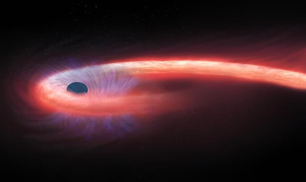
TDEs are astronomical phenomena that occur when a star passes close enough to a supermassive black hole and is pulled apart by the black hole’s tidal forces, causing the process of disruption. Such tidally-disrupted stellar debris starts raining down on the black hole and radiation emerges from the innermost region of accreting debris, which is an indicator of the presence of a TDE. All in all, the debris stream-stream collision causes an energy dissipation, which may lead to the formation of an accretion disk.
Therefore, TDEs are perceived by astronomers as potentially important probes of strong gravity and accretion physics, providing answers about the formation and evolution of supermassive black holes.