Toggle light / dark theme

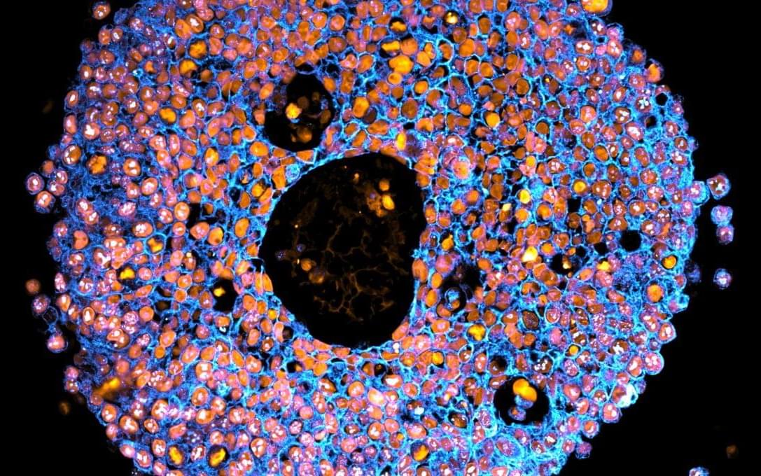
A new kind of microscope called ELVIS is heading to the International Space Station to change how we study life in space. By creating stunning 3D holograms of cells, it allows scientists to observe how organisms adapt to microgravity and other extreme conditions. This could help us understand whe

In 2021, research led by Ryan Flynn, MD, Ph.D., and his mentor, Nobel laureate Carolyn Bertozzi, Ph.D., opened a new chapter in biology, characterizing a new kind of player on the cell surface: glycoRNAs. Extending this discovery recently in Cell, Flynn and colleagues showed that glycoRNAs form highly organized clusters with RNA-binding proteins on the cell surface. These clusters appear to regulate communication between cells and their environment.

Now, reporting in Nature Biotechnology, Flynn’s team in the Stem Cell Program at Boston Children’s Hospital and Dana-Farber/Boston Children’s Cancer and Blood Disorders Center demonstrate a first application of this newfound biology: fighting cancer.

With collaborators at the Cambridge (UK) Stem Cell Institute, led by Konstantinos Tzelepis, Ph.D., and Maria Eleftheriou, Ph.D., they show that one RNA-binding protein on the cell surface, NPM1, could be a potent, selective target for treating (AML), as well as .

Suppose you want to make a tiny robot to perform surgery inside a human patient. To avoid damaging healthy tissue and to squeeze into tight spots, the robot should be squishy. And manipulating the robot’s movements with magnetic fields would make sense, as tissues don’t respond to magnetism. But what material would you use for the robot’s limbs? Magnetic materials are stiff and brittle. Embedding tiny particles of them in a rubbery matrix could work, but the thinner—and therefore bendier—you make the composite material, the less it responds to a magnetic field. Heinrich Jaeger of the University of Chicago, Monica Olvera de la Cruz of Northwestern University, Illinois, and their collaborators have now overcome that obstacle by making thin, flexible sheets out of self-assembled nanoparticles of magnetite [1]. Even a modest field of 100 milliteslas can lift a sheet and bend it by 50°, they found.

At room temperature, magnetite (Fe3O4) is ferrimagnetic—that is, the magnetic moments in its two sublattices align in opposite directions but with unequal magnitudes, yielding a net magnetization. The smaller a ferrimagnet, the greater the chance that it has a single domain, and therefore the lower the temperature at which the domain’s magnetization will flip. When the sample size gets down to a few tens of nanometers, a ferrimagnet made of randomly flipping particles becomes, in effect, a paramagnet—that is, it lacks a net magnetization and is attracted by an applied magnetic field. The attraction can be strong. The discoverers of this phenomenon in 1959 dubbed it superparamagnetism [2].

The researchers realized that a sheet made from a single layer of superparamagnetic particles could serve as a viable material for the magnetic actuation of small soft robots. To create the layers, they suspended magnetite nanoparticles in droplets of water coated with an organic solvent. The solvent attracted the nanoparticles, which migrated to a droplet’s surface. The water slowly evaporated, leaving behind a layer of closely packed nanoparticles draped on the droplet’s support structure, a square copper grid. Each of the 20 × 20 µm squares supported a single sheet. As shown in the figure, some of the sheets happened to have a single unattached corner.

A device made of multilayer graphene exhibits topologically protected edge currents whose direction can be switched using an electric field.

Topological phases of matter have captivated physicists for several decades, promising exotic phenomena and new paradigms for electronic devices [1]. So-called Chern insulators—systems exhibiting quantized Hall conductance without an external magnetic field—are particularly enticing. These materials support dissipationless, one-way electron transport along their edges, which could enable robust low-power electronics or even form the backbone of future topological quantum-computing architectures [2]. Yet, the defining feature of a Chern insulator—its chirality, which determines the direction of the edge-state current—is set by material symmetry and is therefore notoriously rigid and difficult to manipulate dynamically [3–5].

A brief episode of anxiety may have a bigger influence on a person’s ability to learn what is safe and what is not. Research recently published in npj Science of Learning has used a virtual reality game that involved picking flowers with bees in some of the blossoms that would sting the participant—simulated by a mild electrical stimulation on the hand.

Researchers worked with 70 neurotypical participants between the ages of 20 and 30. Claire Marino, a research assistant in the ZVR Lab, and Pavel Rjabtsenkov, a Neuroscience graduate student at the University of Rochester School of Medicine and Dentistry, were co-first authors of the study.

Their team found that the people who learned to distinguish between the safe and dangerous areas—where the bees were and were not—showed better spatial memory and had lower , while participants who did not learn the different areas had higher anxiety and heightened fear even in safe areas.

When a planet’s orbit brings it between Earth and a distant star, it’s more than just a cosmic game of hide and seek. It’s an opportunity for NASA to improve its understanding of that planet’s atmosphere and rings. Planetary scientists call it a stellar occultation and that’s exactly what happened with Uranus on April 7.

Observing the alignment allows NASA scientists to measure the temperatures and composition of Uranus’s stratosphere—the middle layer of a planet’s atmosphere—and determine how it has changed over the last 30 years since Uranus’s last significant occultation.

“Uranus passed in front of a star that is about 400 light years from Earth,” said William Saunders, planetary scientist at NASA’s Langley Research Center in Hampton, Virginia, and science principal investigator and analysis lead, for what NASA’s team calls the Uranus Stellar Occultation Campaign 2025.

For biologists, seeing is believing. But sometimes biologists have a hard time seeing. One particularly vexing challenge is seeing all the molecules in an intact tissue sample, down to the level of single cells, simultaneously. Detecting the location of hundreds or thousands of biomolecules—from lipids to metabolites to proteins—in their native environment allows researchers to better understand their functions and interactions. Unfortunately, scientists don’t have great tools to accomplish this task.

Now a multi-institutional research team has developed a tissue expansion method that enables scientists to use imaging to simultaneously detect hundreds of molecules at the single cell level in their native locations. Their paper is published in the journal Nature Methods.

Imaging methods, including most types of microscopy, provide a view of molecules inside cells. But they can track only a select handful of molecules at one time, and they can’t detect all types of biomolecules, including some lipids. Other methods, like regular mass spectrometry, can detect hundreds of molecules but don’t work on intact samples, so researchers can’t see how the biomolecules are oriented.

A new comparison and analysis of the genomes of species in the genus Malus, which includes the domesticated apple and its wild relatives, revealed the evolutionary relationships among the species and how their genomes have evolved over the past nearly 60 million years.

The research team identified structural variations among the genomes and developed methods for identifying genes associated with desirable traits, like tastiness and resistance to disease and cold, that could help guide future breeding programs.

A paper describing the research, conducted by an international team that includes Penn State biologists, was published in the journal Nature Genetics.

Researchers from the Institute of Modern Physics (IMP) of the Chinese Academy of Sciences (CAS) have proposed a key indicator that may reveal the emergence of quark-gluon plasma (QGP) by analyzing particle “fingerprints” generated in heavy-ion collisions.

Published in Physics Letters B, the study provides a new perspective for exploring the evolution of matter in the .

About 13.8 billion years ago, within a millionth of a second after the Big Bang, the universe existed in an ultra-hot and dense state. Instead of protons and neutrons, the fundamental building blocks of matter were free quarks and gluons—a unique state known as QGP. As the universe expanded and cooled, the QGP gradually condensed into the we recognize today.