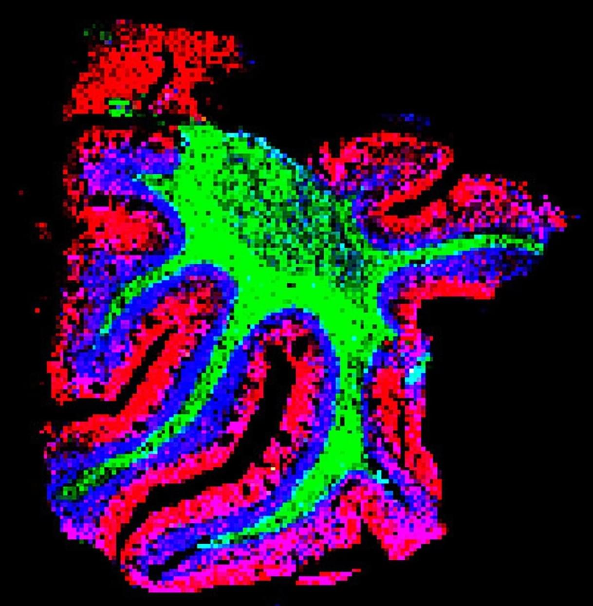
For biologists, seeing is believing. But sometimes biologists have a hard time seeing. One particularly vexing challenge is seeing all the molecules in an intact tissue sample, down to the level of single cells, simultaneously. Detecting the location of hundreds or thousands of biomolecules—from lipids to metabolites to proteins—in their native environment allows researchers to better understand their functions and interactions. Unfortunately, scientists don’t have great tools to accomplish this task.
Now a multi-institutional research team has developed a tissue expansion method that enables scientists to use mass spectrometry imaging to simultaneously detect hundreds of molecules at the single cell level in their native locations. Their paper is published in the journal Nature Methods.
Imaging methods, including most types of microscopy, provide a view of molecules inside cells. But they can track only a select handful of molecules at one time, and they can’t detect all types of biomolecules, including some lipids. Other methods, like regular mass spectrometry, can detect hundreds of molecules but don’t work on intact samples, so researchers can’t see how the biomolecules are oriented.