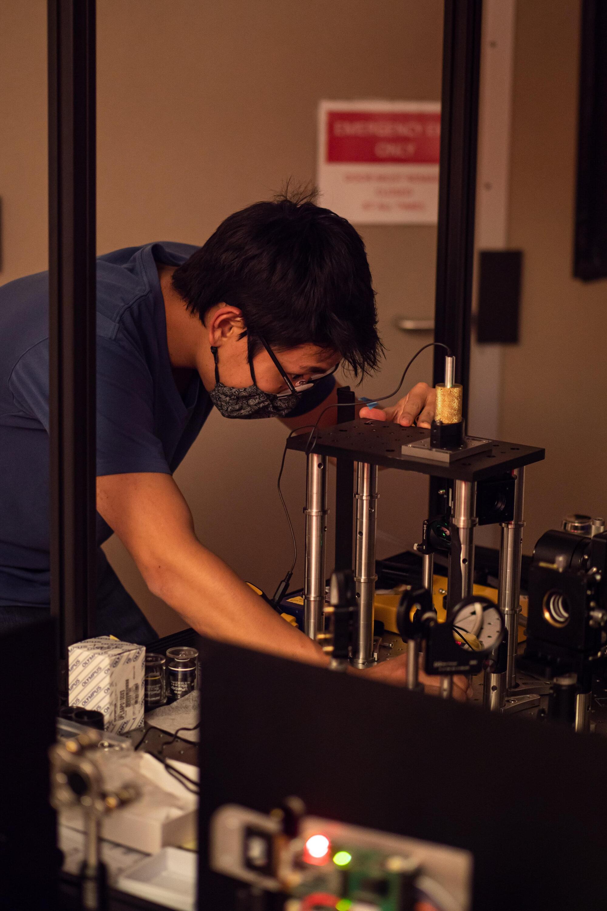
Researchers have developed a high-speed 3D imaging microscope that can capture detailed cell dynamics of an entire small whole organism at once. The ability to image 3D changes in real time over a large field of view could lead to new insights in developmental biology and neuroscience.
“Traditional microscopes are constrained by how quickly they can refocus or scan through different depths, which makes it difficult to capture fast, 3D biological processes without distortion or missing information,” said Eduardo Hirata Miyasaki, who performed the work while in Sara Abrahamsson’s lab at the University of California Santa Cruz (UCSC) and is now at the Chan Zuckerberg Biohub.
“Our new system extends the multifocus microscopy (MFM) technique Abrahamsson developed by using a 25-camera array to push the limits of speed and volumetric imaging. This leap in efficiency opens the door to studying small living systems in motion without disrupting them.”


