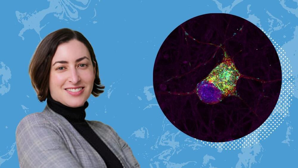
Sarah Cohen and two others will pioneer a method to view these molecular structures and their interactions in live stem cells.

In this interview, I sit down with Simon Critchley, Hans Jonas Professor of Philosophy at The New School for Social Research in New York, to explore his provocative new book, On Mysticism. Drawing on medieval Christian figures like Julian of Norwich and Marguerite Porete, Critchley argues that ecstatic experience, intense love, and a willingness to be “outside oneself” can offer a counterbalance to the narrowly rational outlook dominant in modern philosophy. Throughout our conversation, he probes the boundaries of faith and reason, discuss the possibility of maintaining mysticism alongside science, and question the role of philosophy itself in shaping our cultural consciousness. What follows is only a short, edited extract from Critchley’s call for more openness, both in our thinking and our collective search for meaning. Link to the full interview.
Sign up to get exclusive access.

Researchers at the University of Oklahoma have developed a breakthrough method of adding a single nitrogen atom to molecules, unlocking new possibilities in drug research and development. Now published in the journal Science, this research is already gaining international attention from drug manufacturers.
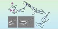
Nitrogen atoms and nitrogen-containing chemical structures, called heterocycles, play a pivotal role in medicinal chemistry and drug development. A team led by OU associate professor Indrajeet Sharma has demonstrated that by using a short-lived chemical called sulfenylnitrene, researchers can insert one nitrogen atom into bioactive molecules and transform them into new pharmacophores that are useful for making drugs.
This process is called skeletal editing and takes inspiration from Sir Derek Barton, the recipient of the 1969 Nobel Prize in Chemistry.
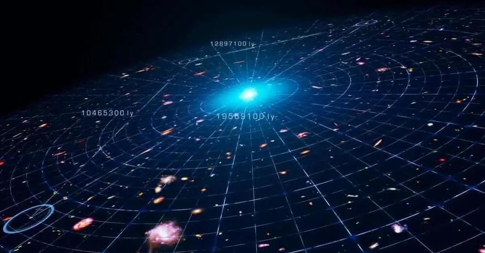
This idea stems from General Relativity, which shows that space and time are not fixed but dynamic and interwoven. Two key discoveries in the early 20th century solidified this understanding. First, Vesto Slipher observed that light from many nebulae was redshifted, indicating they were moving away. Second, Edwin Hubble measured distances to these galaxies and found that the farther they were, the faster they receded. This correlation, now known as Hubble’s Law, confirmed that the Universe is expanding.
Scientists often use analogies to explain this phenomenon. The “balloon analogy” imagines galaxies as coins on a balloon’s surface, moving apart as the balloon inflates. Another analogy is a loaf of raisin bread dough, where the raisins (galaxies) move apart as the dough (space) expands. However, these analogies fall short in some respects. Unlike the dough or balloon, the Universe doesn’t expand into anything; it’s all there is.
Observations suggest the observable Universe is only a fraction of a potentially infinite cosmos. While light from unseen regions will eventually reach us, expanding spacetime itself ensures galaxies continue moving farther apart. The theory of cosmic inflation suggests that our Universe is one “bubble” in a vast multiverse, though these regions remain isolated from one another.

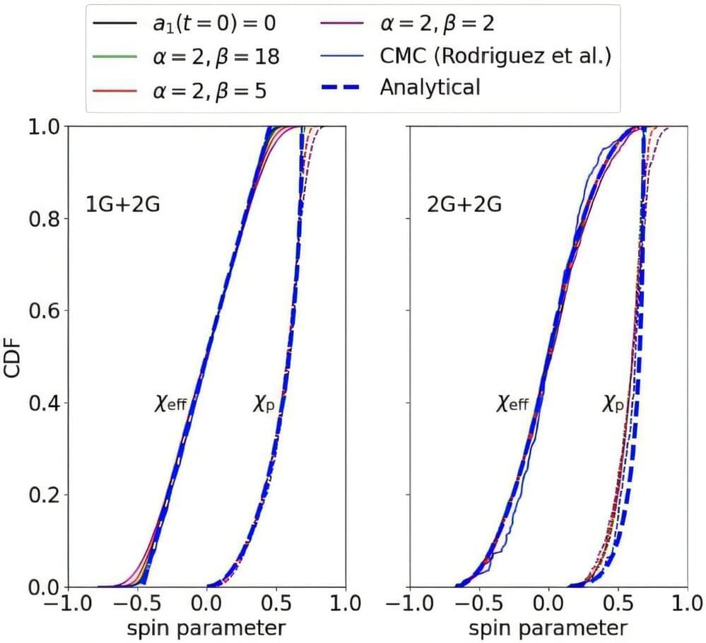
The size and spin of black holes can reveal important information about how and where they formed, according to new research.
The study, led by scientists at Cardiff University, tests the idea that many of the black holes observed by astronomers have merged multiple times within densely populated environments containing millions of stars.
The work is published in the journal Physical Review Letters.