Toggle light / dark theme

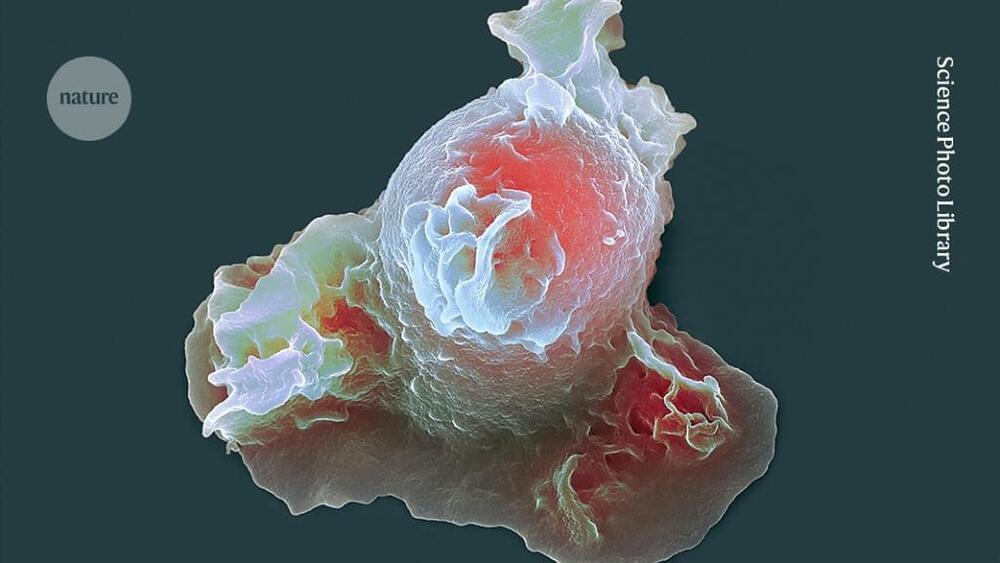
For years, there has been a long-held belief that acute viral infections like Zika or COVID-19 are directly responsible for neurological damage, but researchers from McMaster University have now discovered that it’s the immune system’s response that is behind it.

The research, published on Feb. 5, 2024, in Nature Communications, was led by Elizabeth Balint, a Ph.D. student at McMaster, and Ali Ashkar, a professor with the Department of Medicine and the Canada Research Chair in Natural Immunity and NK Cell Function.

“We were interested in trying to understand why so many are associated with ,” says Balint. “Our evidence suggests that it’s not the virus itself that causes the damage, but a unique population of T cells, which are part of the immune system, that are actually responsible for the damage.”

Acetamiprid-induced oxidative stress can harm DNA and tRNA, leading to health problems. A study conducted by Huixia Zhang at Macau University of Science and Technology in 2023 introduced a comprehensive approach to assessing acetamiprid-induced oxidative damage to tRNA in human cells through oxidized nucleotide and tRNA profiling. Acetamiprid, a modern insecticide, is known for causing oxidative stress and related toxicity. Despite its impact on oxidative stress, the effects of acetamiprid-induced oxidative stress on RNA, especially tRNA, remained unexplored until this study.

Acetamiprid was found to elevate reactive oxygen species (ROS) production in HepG2 and LO2 cells, contributing to mitochondrial damage, free radical generation, and antioxidant status depletion. Oxidative damage to DNA and RNA can harm organisms, with prior research addressing RNA damage in aging, neurodegenerative diseases, and mental illnesses. However, its role in acetamiprid-induced toxicities has not been investigated.

The study employed TMSD labeling-based LC-MS/MS to measure oxidized nucleotide levels in HepG2 and LO2 cells treated with two mM acetamiprid. It also examined the impact of acetamiprid on the 8-oxo-G content of tRNAs and created volcano plots to compare RNase T1 digestion products of tRNAs from untreated and acetamiprid-treated cells.

Researchers have not only built an AI child, but are now training AI using headcam footage from a human baby as well.

In a press release, New York University announced that its data science researchers had strapped a camera to the head of a real, live, human toddler for 18 months to see how much an AI model could learn from it.

Most large language models (LLMs), like OpenAI’s GPT-4 and its competitors, are trained on “astronomical amounts of language input” that are many times larger than what infants receive when learning to speak a language during the first years of their lives.

A team of scientists at the University of Wisconsin-Madison claim to have 3D-printed functional human brain tissue for the first time.

They hope their research could open the doors for the development of treatments for existing neurological disorders, including Alzheimer’s and Parkinson’s disease.

As detailed in a new paper published in the journal Cell Stem Cell, the team flipped the usual method of 3D-printing on its side, fabricating horizontal layers of brain cells encased in soft “bio-ink” gel.