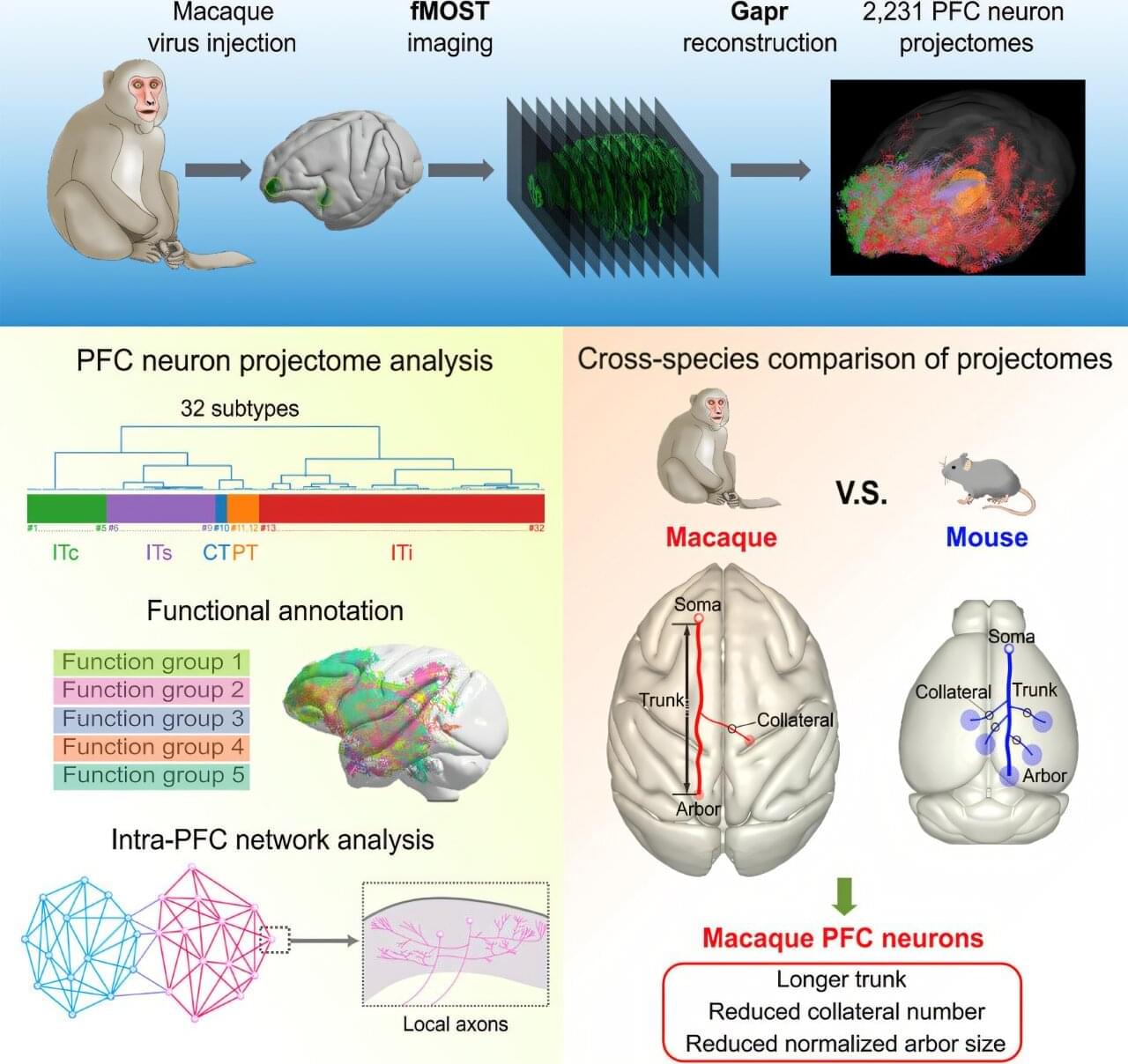
In a study published in Cell on July 10, researchers reported the first comprehensive study of whole-brain projectomes of the macaque prefrontal cortex (PFC) at the single-neuron level and revealed the organization of macaque PFC connectivity.
The team from the Center for Excellence in Brain Science and Intelligence Technology (CEBSIT) of the Chinese Academy of Sciences, along with a team from the HUST-Suzhou Institute for Brainsmatics, compared macaque and mouse PFC single-neuron projectomes and revealed highly refined axon targeting and arborization in primates.
The PFC in primates, including humans, has dramatically expanded over the course of evolution, which is believed to be the structural basis of high cognitive functions. Previous studies of PFC connectivity in non-human primates have mainly relied on population-level viral tracing and functional magnetic resonance imaging (fMRI), which in general lack single-cell resolution to examine projection diversity. Meanwhile, whole-brain imaging data for tracing axons in the primate brain are massive in size.