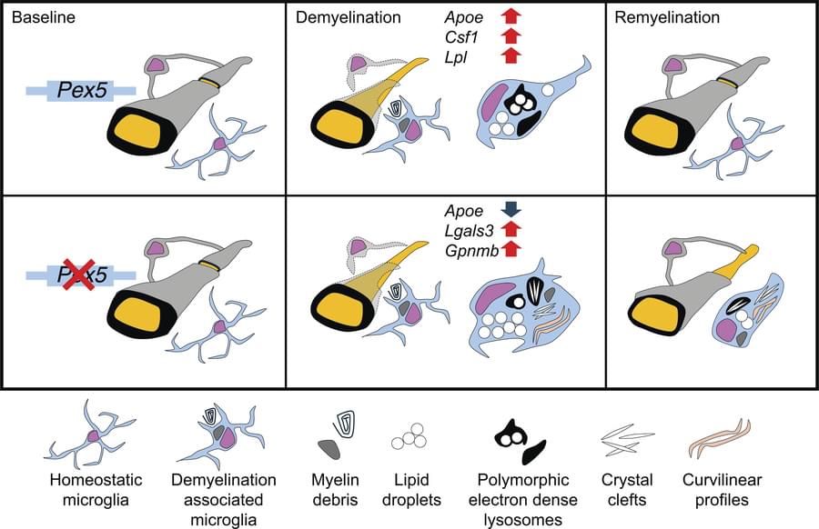
Jian Hu & team use mouse models to show peroxisomes license myelin debris degradation in myeloid cells, which enables debris clearance and remyelination after myelin damage:
The figure shows TEM micrographs of phagocytes in which PEX5 loss (bottom panel) aggregates lipid droplets and crystal accumulation.
1Department of Cancer Biology, MD Anderson Cancer Center, Houston, Texas, USA.
2University of Texas MD Anderson Cancer Center UTHealth Graduate School of Biomedical Sciences, Houston, Texas, USA.
3University of Puerto Rico School of Medicine, San Juan, Puerto Rico.