Toggle light / dark theme

A POSSIBLE cure for inherited eye diseases which cause blindness is within sight thanks to a breakthrough recorded by a Tasmanian-led research team.

Associate Professor Alex Hewitt, an ophthalmologist and a researcher with the University of Tasmania’s Menzies Institute for Medical Research and the School of Medicine, said his team had successfully edited adult eye tissue genes in the laboratory to replace unwanted genes that caused blindness.

Dr Hewitt said he was optimistic Tasmanian doctors could soon start wielding molecular gene shears inside the eyes of humans.

Read more

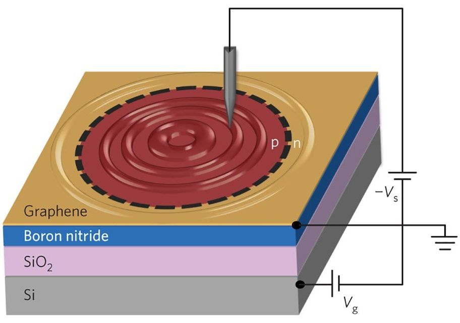
(Phys.org)—A team of researchers with the University of California, MIT, Lawrence Berkeley National Laboratory and the National Institute for Materials Science in Japan has created images of relativistic electrons trapped in graphene quantum dots. In their paper published in the journal Nature Physics the team describes how they achieved this feat and where they plan to take their work in the future.

As the many unique properties of graphene continue to unfold, scientists seek new ways to harness and eventually make use of them. One such use might be to control electrons to allow their use in nano-scaled devices, which could also inadvertently lead to a deeper understanding of Dirac fermions. In this new effort, the researchers have made progress in that area by devising a means for capturing and holding electrons and for creating images of the result.

Obtaining images of electron waveforms has thus far been particularly difficult—virtually all existing methods have resulted in too many defects. To get around such problems, the researchers took another approach to capturing the electrons. They first created circular p-n junctions by sending voltage through the tip of a scanning tunneling microscope down to a graphene sample below. At the same time, they also applied voltage to a slab of silicon underneath the piece of graphene, which was kept separated by a layer of silicon-oxide and a flake of . Doing so caused defects in the boron nitride to ionize, resulting in charges migrating to the graphene.

Read more

Quantum physics: The human body is controlled by electrical impulses in, for example, the brain, the heart and nervous system. These electrical signals create tiny magnetic fields, which doctors could use to diagnose various diseases, for example diseases of the brain or heart problems in young foetuses. Researchers from the Niels Bohr Institute have now succeeded in developing a method for extremely precise measurements of such ultra-small magnetic fields with an optical magnetic field sensor. The results are published in the scientific journal, Scientific Reports.

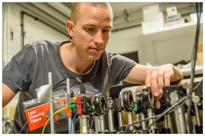
Assistant Professor Kasper Jensen in the Quantop research group’s laboratories at the Niels Bohr Institute where the experiments are carried out. (Photo: Ola Jakup Joensen)

Small magnetic fields from the human body can usually only be picked up by very sensitive superconducting magnetic field sensors that have to be cooled by liquid helium to near absolute zero (which is minus 273 degrees Celsius). But now researchers from the Niels Bohr Institute at the University of Copenhagen have developed a much cheaper and more practical optical magnetic field sensor that even works at room temperature or at body temperature.

Read more

Scientists found that quantum fingerprinting protocol can surpass the classical limit for solving communication complexity problems.

A new study has experimentally demonstrated a quantum fingerprinting protocol and shown that it can surpass the classical limit for solving communication complexity problems. Scientists say that in these problems, two parties each have a message, and they both share some of their message with a referee, who has to decide whether the two messages are the same or not. The classical limit requires that a minimum amount of information must be transmitted between each party and the referee in order for the referee to make this decision.

Researchers found that the best communication complexity protocols require transmission of data that is two orders of magnitude larger than the classical limit.

Read more

By Dr. Robert Green, postdoctoral fellow, Quantum Matter Institute

In the field of quantum matter research, we seek to uncover materials with properties that may find applications in new technologies. My team and I study the properties of various materials at an atomic level to find innovative ways that they can be used to compose the next generation of computer chips. Our research results in large amounts of experimental data. One of the toughest challenges is to analyze and present the data in a meaningful way, for not only our understanding of their underlying complex, quantum principles, but also for wider audiences, including fellow researchers in the field.

One of our key research projects aims to uncover properties in materials that might be used to make smaller, more energy efficient computer chips — five to 10 years from now. In accordance with Moore’s Law, the number of transistors and overall processing power within a chip has doubled every two years for over four decades. But as chips have become more and more powerful, technological demands also continue to expand and the devices that use these chips are also becoming more portable. As a result, conventional practices of making chips are straining the laws of physics to incorporate more transistors within a shrinking area.

Read more
High-performance detectors that are compatible with mainstream semiconductor device fabrication deliver high speed, ultra-sensitivity, and good timing resolution.

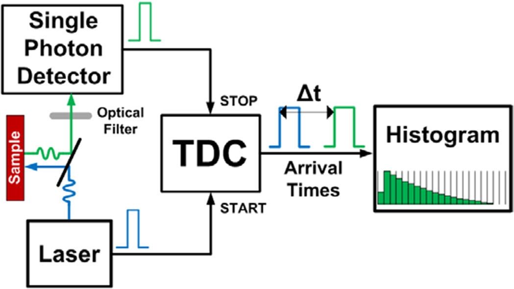
Recent advances in biomedical imaging include the enhancement of image contrast, 3D sectioning capability, and compatibility with specialized imaging modes such as fluorescence lifetime imaging (FLIM).1–3 Compared with other imaging methods, FLIM offers the highest image contrast because it measures the lifetime of the fluorescence, rather than just its intensity or wavelength characteristics. The contrasting fluorescence lifetime attributes can then enable the observer to discriminate between regions, such as identifying healthy and diseased tissue for cancer detection. In conventional FLIM, a discrete single-photon detector, typically based on photomultiplier tube (PMT) technology, enables the acquisition of a single focal spot.4 This focal spot is then raster-scanned across the field of view to form an image. This approach, however, requires sequential scanning—pixel by pixel—and thus results in a slow image acquisition rate.

Read more

Human brain is made up of a billion nerve cells called neurons and various other types of cells and is the most complex machine ever known. Even after years of research and studies we still do not have a complete understanding of how it works — how it controls every single thing we ever do. In order to unravel one such mysteries of the brain, researchers at the Carnegie Mellon University set out to find out why brain makes mistakes. The study was conducted as part of Carnegie Mellon’s BrainHub, a university initiative that focuses on how the structure and activity of the brain give rise to complex behaviors.

Brain

Read more