Antarctica’s native microorganisms are a hearty bunch, able to eke out a living on the planet’s coldest, highest, driest, windiest and emptiest continent. But the region wasn’t always quite as hospitable as it is today.
Photo Credit: Byron Adams

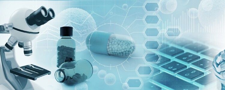
There are lots of exciting companies working in the aging field, and it’s a great time to tell you about some of the more interesting ones. Most of these companies are a while away from human trials yet, but their innovations could possibly be truly game changing.
Underdog Pharmaceuticals is a spin-off company of SENS Research Foundation and is developing a novel approach to treating atherosclerosis.
Atherosclerosis is the number one killer worldwide, and it currently has no totally effective solution. There are three ways in which current medicine tries to address it: Lifestyle changes, including diet and exercise; drugs that slow down the rate of cholesterol accumulation; and interventions such as stents and bypass surgery.
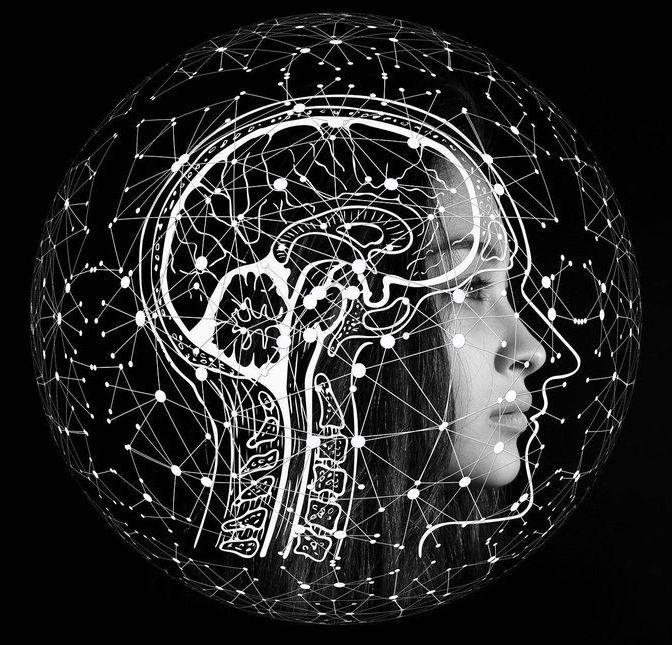
In neurological diseases such as Alzheimer’s disease, Parkinson’s disease or Multiple Sclerosis, brain neurons are constantly being lost, resulting in memory lapses, speech disorders, mood swings and movement disorders, for example, as well as muscle tremors in the case of Parkinson’s. After six years of development, MedUni Vienna researchers from the Department of Neurology (Head: Thomas Berger), led by Roland Beisteiner, have developed a new method of treatment that represents a world first. Using a non-invasive ultrasound technique, it is now possible to reach all areas of the brain and activate neurons that can help to regenerate brain functions. The preliminary data, which have been prominently published on the international stage, show that this can improve brain performance. This has positioned Vienna as a world leader in an important sector of medicine.
The new method is called transcranial pulse stimulation with ultrasound (TPS) and was developed in collaboration with Swiss commercial partner Storz Medical and its project leader, Ernst Marlinghaus. “For the first time in the world, TPS enables us to penetrate into all areas of the brain by means of an ultrasound pulse delivered directly to the skull in a non-invasive, painless procedure, during which the patient is fully conscious, and to specifically target particular areas of the brain and stimulate them,” explains Beisteiner. The study was part of the inter-university cluster led by Roland Beisteiner and Tecumseh Fitch, which is attempting to improve patients’ brain functions by means of brain stimulation and is being jointly run by MedUni Vienna and the University of Vienna. Such clinical procedures must be carried out with great precision and must be tailored to the individual patient. However, the existing electromagnetic techniques such as e.g.
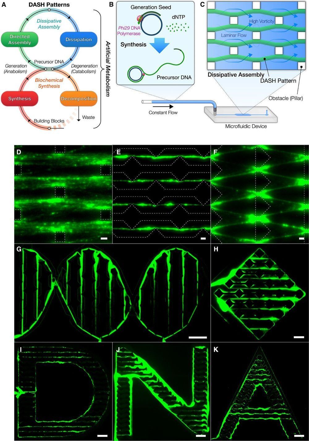
Interesting research paper on a new nanobot technology. I’m watching for ways in which suitable substrates for mind uploading can be constructed, and DNA self-guided assembly has potential.
Here are some excerpts and a weblink to the paper:
“…Chemical approaches have opened synthetic routes to build dynamic materials from scratch using chemical reactions, ultimately allowing flexibility in design…”
… As a realization of this concept, we engineered a mechanism termed DASH—DNA-based Assembly and Synthesis of Hierarchical materials—providing a mesoscale approach to create dynamic materials from biomolecular building blocks using artificial metabolism. DASH was developed on the basis of nanotechnology that uses DNA as a generic material ranging from nanostructures to hydrogels, for enzymatic substrates, and as linkers between nanoparticles…”
“…Next, to illustrate the potential uses of self-generated materials, we created various hybrid functional materials from the DASH patterns. The DASH patterns served as a versatile mesoscale scaffold for a diverse range of functional nanomaterials beyond DNA, ranging from proteins to inorganic nanoparticles, such as avidin, quantum dots, and DNA-conjugated gold nanoparticles (AuNPs) (Fig. 4D, figs. S37 and S38, and Supplementary Text). The generated patterns were also rendered functional with catalytic activity when conjugated with enzymes (figs. S39 and S40 and Supplementary Text). We also showed that the DNA molecules within the DASH patterns retained the DNA’s genetic properties and that, in a cell-free fashion, the materials themselves successfully produced green fluorescent proteins (GFPs) by incorporating a reporter gene for sfGFP (Fig. 4E and figs. S9 and S41) (40). The protein production capability of the materials established the foundation for future cell-free production of proteins, including enzymes, in a spatiotemporally controlled manner.
…” Our implementation of the concept, DASH, successfully demonstrated various applications of the material. We succeeded in constructing machines from this novel dynamic biomaterial with emergent regeneration, locomotion, and racing behaviors by programming them as a series of FSAs. Bottom-up design based on bioengineering foundations without restrictions of life fundamentally allowed these active and programmable behaviors. It is not difficult to envision that the material could be integrated as a locomotive ele-ment in biomolecular machines and robots. The DASH patterns could be easily recognized by naked eyes or smartphones, which may lead to better detection technologies that are more feasible in point-of-care settings. DASH may also be used as a template for other materials, for example, to create dynamic waves of protein expression or nanoparticle assemblies. In addition, we envision that further expansion of artificial metabolism may be used for self-sustaining structural components and self-adapting substrates for chemical production pathways. Ultimately, our material may allow the construction of self-reproducing machines through the production of enzymes from generated materials that, in turn, reproduce the material. Our biomaterial powered by artificial metabolism is an important step toward the creation of “artificial” biological systems with dynamic, life-like capabilities.”…

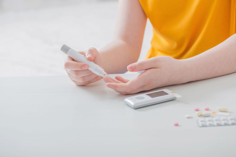
The insulin pumps diabetics currently rely on do a great job of delivering the hormone as needed, but need regular replacing due to what are known as fibrils. These form over a day or two as insulin compounds accumulate into clumps and create the risk of blockages, but scientists in Australia have engineered what they say is a safer alternative, with egg yolks serving as their starting point.
The formation of fibrils means that diabetics need to replace their insulin pumps every 24 to 72 hours to avoid the risk of dangerous blockages, which bring with them a risk of life-threatening under-dosing. Beyond the dangers to the patient’s well-being, the need to regularly replace the pump increases the workload needed to manage their disease and means that portions of the medicine often go to waste.
So, there is considerable interest in developing synthetic insulin that doesn’t behave in this way. Researchers at Melbourne’s Florey Institute of Neuroscience and Mental Health approached this problem through a new technique it developed with scientists in Japan, whereby the insulin is engineered from egg yolks to allow for greater freedom over the final design.

Scientists have discovered that when an essential key protein needed to generate novel brain cells during pregnancy and early childhood days of the offspring is missing, which makes the brain goes haywire. This particular deprivation causes an imbalance in brain’s circuitry can lead to long-term cognitive and movement behaviours characteristic of autism spectrum disorder.” During brain development, there is a coordinated series of events that have to occur at the right time and the right place in order to establish the appropriate number of cells with the right connections,” said Juan Pablo Zanin, Rutgers-Newark research associate, and lead author.” Each of these steps is carefully regulated and if any of these steps are not regulated correctly, this can impact behaviour.
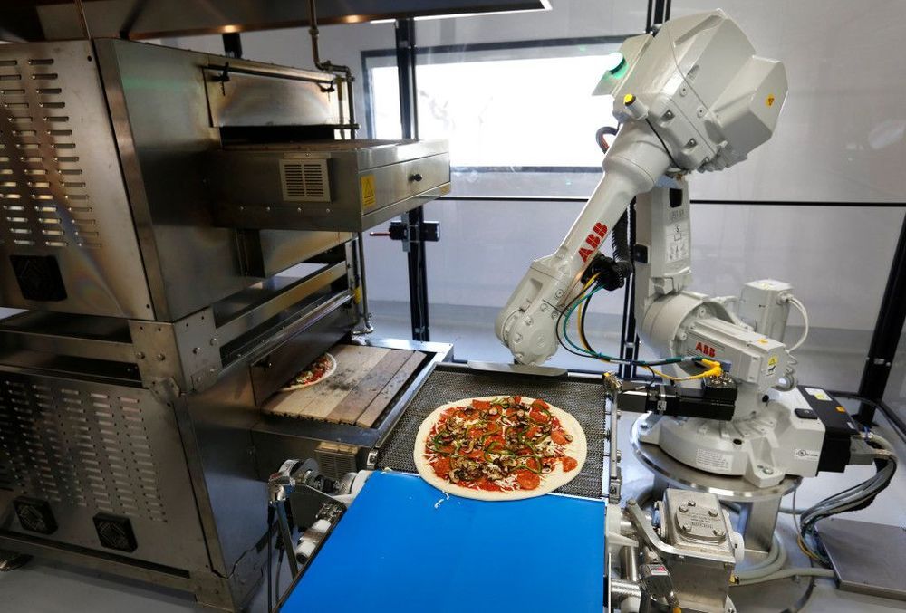
Zume Pizza, the Mountain View company that used robots to make its pizzas, has made its last delivery.
In filings with the state Employment Development Department, Zume said it is cutting 172 jobs in Mountain View, and eliminating another 80 jobs at its facility in San Francisco. Zume Chief Executive Alex Garden made the annoucement about Zume in an email to company employees on Wednesday.
“With admiration and sadness, we are closing Zume Pizza today,” Garden said in his email “Over the last four years this business has been our invention test bed and has been our inspiration for many of the growth businesses we have at Zume today.”