Toggle light / dark theme

Huge unveiling of schizophrenia brain cells show new treatment targets

If you thought it was easy to analyze brain cells, think again.

When you take a brain tissue sample, all that your analysis would normally show you is an average for all the present. And since there are a whole lot of cell types in our brain— and others—you’ll get a sort of cell smoothie, which makes it difficult if not impossible to tell the cells apart, let alone study them.

It is like wanting to know how many green M&M’s there are in a bowl, but instead just getting told how many colors there are. You are not really getting the answer you wanted.

A telltale protein spreads throughout the brain in distinct patterns based on patients’ Alzheimer’s phenotype

New imaging of patients with Alzheimer’s demonstrates how a telltale protein spreads throughout the brain based on the phenotype of the disease, i.e., whether the condition is dominated by forgetfulness, or atrophy in a specific brain region. The research offers a host of illuminating clues that ultimately may inform new treatment strategies.

The protein is known as tau and a large multi-disciplinary team of brain researchers at McGill University in Montreal has been able to trace the protein’s patterns in living patients via magnetic resonance imaging (MRI). Alzheimer’s disease is intimately linked to tau, which can form tangles in the brain that irrevocably damage neurons.

The patterns detected by McGill scientists apparently are unique to the phenotype of Alzheimer’s afflicting the patient. This staggering finding opens an intriguing new window into the molecular mechanisms of the disease. And while many features of Alzheimer’s are the same from one patient to the next, phenotypes are a hallmark of the condition. Tracking tau patterns is a specialty of the scientists at McGill, who found that the intrinsic connectivity of the human brain itself provides the scaffolding for the aggregation of tau in distinct variants of the disease.

Neuroimaging study reveals functional and structural brain abnormalities in people with post-treatment Lyme disease

In a study using specialized imaging techniques, Johns Hopkins Medicine researchers report distinctive changes in the “white matter” and other brain tissue physiology of those with post-treatment Lyme disease, a condition affecting 10% to 20% of the nearly half a million Americans who contract Lyme disease annually.

The study’s findings, published October 26 in the journal PLOS ONE, substantiate and help validate that memory difficulties and other cognitive difficulties experienced long-term by individuals with post-treatment Lyme disease are linked to functional and structural changes in the brain.

Lyme disease, whose early symptoms may include a characteristic rash, flu-like aches and fever, , and fatigue, is treated using a rigorous course of antibiotics, which usually clears the illness.

‘Time Cells’ in The Human Brain Encode The Flow of Time, Scientists Say

How does the human brain keep track of the order of events in a sequence?

Research suggests that ‘time cells’ – neurons in the hippocampus thought to represent temporal information – could be the glue that sticks our memories together in the right sequence so that we can properly recall the correct order in which things happened.

Evidence for these kinds of sequence-tracking time cells has previously been found in rats, where specific neuron assemblies are thought to support the recollection of events and the planning of action sequences – but for a long time, less was known about how episodic memory is encoded in the human brain.

A new study reveals how word and face recognition can be supported with only half the brain

Researchers conclude that one hemisphere of the brain can adequately function as if it were doing so for two hemispheres.

People who underwent surgery as children to remove half of their brain were still able to accurately recognize differences between pairs of words or faces.

The research was done to study brain plasticity and perception. Plasticity is when the brain can be molded to reorganize itself in the hemispheric region not injured, or in this case, the only hemispheric region that is there. The participants were able to correctly identify differences between words or faces with more than 80% accuracy.

This thumb-sized microscope captures ‘neural landscapes’ from deep inside animal brains

The new design came with three fundamental improvements.

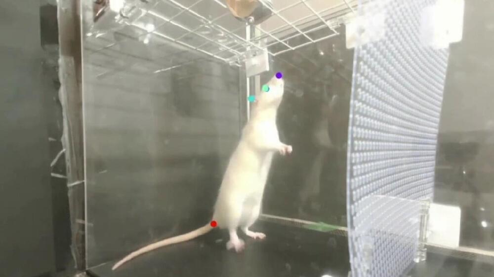
Researchers have finally managed to reduce the two-photon fluorescence microscope into a thumb size device that allows them to see inside the brain of live and active animals. The device called Mini2P weighs just 2.4 grams and can be attached to a mouse’s head without compromising its natural movements.

The microscope can record live images of neural landscapes, the likes of which have never been seen before. The innovation “opens the door to lines of scientific inquiry that were difficult, if not impossible, to initiate,” says Denise Cai, a neuroscientist at the Icahn School of Medicine at Mount Sinai in New York City. feat was achieved by Edvard Moser, professor of Psychology and Neuroscience at the Kavli Institute for Systems Neuroscience, together with Weijing Zong, a biological engineer and neuroscientist at the Moser Group.

/* */