Toggle light / dark theme

Scientists have identified a class of drugs that may have potential to treat a rare and deadly form of brain cancer that affects young children.

The research team, led by Ranjit Bindra, MD, PhD, and colleagues at the Yale Cancer Center, also included co-senior authors Charles Brenner, PhD, professor and DEO of biochemistry at the University of Iowa Carver College of Medicine, and Michael E. Berens, PhD, from the Translational Genomics Research Institute in Phoenix.

The findings, published Aug. 22 in Nature Communications, focus on Diffuse Intrinsic Pontine Glioma (DIPG), a rare, incurable cancer that affects the brainstem in children under age 10. Previous work had identified mutations in a gene called PPM1D as a cause of this cancer.

Nootropics are colloquially known as “smart drugs” – substances that increase cognitive function in healthy people. In this video, bestselling author Dave Asprey discusses two naturally occurring nootropics: caffeine and nicotine.

Microdosing one milligram of the latter — about 5 to 10 percent of a cigarette’s worth — may even protect against Alzheimer’s.

ME Conferences is organizing “18th International Conference on Gastroenterology and Digestive Disorders” during September 16–17, 2019, Dubai, UAE. Conference highlights the theme- Improving Access in Gastroenterology: From Past to Future.

Gastro Meet 2019 is a leading platform for a focused agenda of the current research in the field of gastroenterology which includes, guest lectures, keynotes, symposiums, workshops, exhibitions, panel discussions, and poster sessions. We invite Gastroenterologists, Hepatologists, General Physicians, Microbiologists, Oncologists, Surgeons, Researchers, Students, and Business delegates to join us at Dubai in August 2018 for the 2-day power packed Gastroenterologists Meet.

September 16 (Monday) — 17 (Tuesday)

Download the FREE PDF:
http://trtrevolution.com/PDF

Learn more about Optimizing Your Health, Reversing Your Aging Process and All Things Testosterone and TOT: http://trtrevolution.com/.

To Get Your FREE Paperback copy of the Amazon Best Selling TRT MANual:(Continental USA Only)
http://trtrevolution.com/book

To Listen to the Audio Version of The TRT MANual
http://amzn.to/2exRnf7

When it comes to age reversal and complex regeneration in humans, a basin of research has been put in front of us. How much do we know about the brain, consciousness, and death reversal? As Big Pharma makes a trillion dollars annually, are they interested in finding cures? What valuable lessons are we learning from evolution?

On this episode, we have an in-depth conversation about exciting developments in biology with Bioquark CEO, Ira Pastor.

Fronteirs offers open access. If you want to read full publications this is the place to do so. #enjoy


Download this month’s new releases including the latest research collections on Tinnitus, Brain Aging, Arboviral Infections, and many more! All eBooks are free to download, share and distribute.

Shape the future of your field — and publish your own eBook — by editing an article collection around your research area. Learn more about Research Topics or submit your suggestion to [email protected]

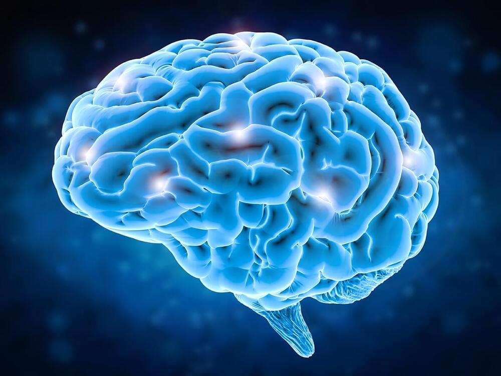
An international team of researchers with partial support from the National Institute of Biomedical Imaging and Bioengineering (NIBIB) developed a new MRI technique that can capture an image of a brain thinking by measuring changes in tissue stiffness. The results show that brain function can be tracked on a time scale of 100 milliseconds – 60 times faster than previous methods. The technique could shed new light on altered neuronal activity in brain diseases.

The human brain responds almost immediately to stimuli, but non-invasive imaging techniques haven’t been able to keep pace with the brain. Currently, several non-invasive brain imaging methods measure brain function, but they all have limitations. Most commonly, clinicians and researchers use functional magnetic resonance imaging (fMRI) to measure brain activity via fluctuations in blood oxygen levels. However, a lot of vital brain activity information is lost using fMRI because blood oxygen levels take about six seconds to respond to a stimulus.

Since the mid-1990s, researchers have been able to generate maps of tissue stiffness using an MRI scanner, with a non-invasive technique called magnetic resonance elastography (MRE). Tissue stiffness can’t be measured directly, so instead researchers use MRE to measure the speed at which mechanical vibrations travel through tissue. Vibrations move faster through stiffer tissues, while vibrations travel through softer tissue more slowly; therefore, tissue stiffness can be determined. MRE is most commonly used to detect the hardening of liver tissue but has more recently been applied to other tissues like the brain.

Magnetic resonance imaging is nothing new, but scientists were able to perform an MRI on a single atom. But how?
» Subscribe to Seeker! http://bit.ly/subscribeseeker
» Watch more Elements! http://bit.ly/ElementsPlaylist

Scientists recently captured the smallest MRI ever while scanning an individual atom. The technique successfully reached a breakthrough level of resolution in the world of microscopy, the detailed MRI can reveal single atoms as well as different types of atoms based on their magnetic interactions.

This breakthrough has potential applications in all kinds of fields, like quantum computing where it could be used to design atomic-scale methods of storing info or when it comes to drug development, the ability to control individual atoms could potentially be used to study how proteins fold and then lead to the development of drugs for diseases like Alzheimers.

In a sense, the researchers combined a version of an MRI machine with a special instrument called a scanning tunneling microscope, which turned out to be a match made in microscopy heaven.

An MRI scanner creates an extremely strong magnetic field around whatever it’s trying to image, temporarily re-aligning the protons in your body with that magnetic field. Then the MRI machine pulses the sample (or patient) with a radiofrequency, which pulls the protons slightly out of alignment with the magnetic field. And after the brief radiofrequency pulse is over, the protons snap back into alignment with the field, and the energy that’s released as the protons move back into place with the magnetic field is what is detected and visualized by the machine.

And a scanning tunneling microscope is used for imaging really tiny surfaces, and it can pick up certain properties like size and molecular structure.