New brain imaging research shows that structural damage in schizophrenia spectrum disorders may begin in specific “epicenter” regions before spreading across connected brain networks.
Every day, our brains transform quick impressions, flashes of inspiration, and painful moments into enduring memories that underpin our sense of self and inform how we navigate the world. But how does the brain decide which bits of information are worth keeping—and how long to hold on?
Now, new research demonstrates that long-term memory is formed by a cascade of molecular “timers” unfolding across brain regions. With a virtual reality-based behavioral model in mice, the scientists discovered that long-term memory is orchestrated by key regulators that either promote memories into progressively more lasting forms or demote them until they are forgotten.
The findings, published in Nature, highlight the roles of multiple brain regions in gradually reorganizing memories into more enduring forms, with gates along the way to assess salience and promote durability.
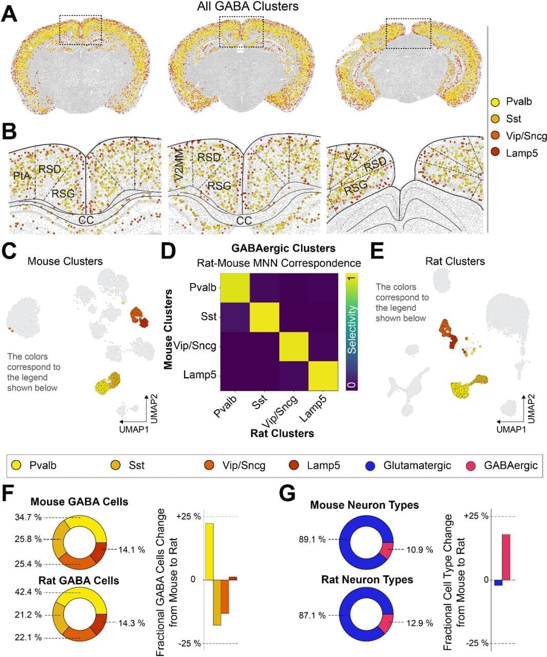
The same brain cells linked to disorientation in Alzheimer’s disease have been preserved—and even slightly increased—across millions of years of evolution.
A new University of Michigan study suggests these neurons are vital to evolutionary survival. Nature has guarded and amplified them through countless generations, helping mammals instinctively know where they are in their environments. The research is published in The Journal of Neuroscience.
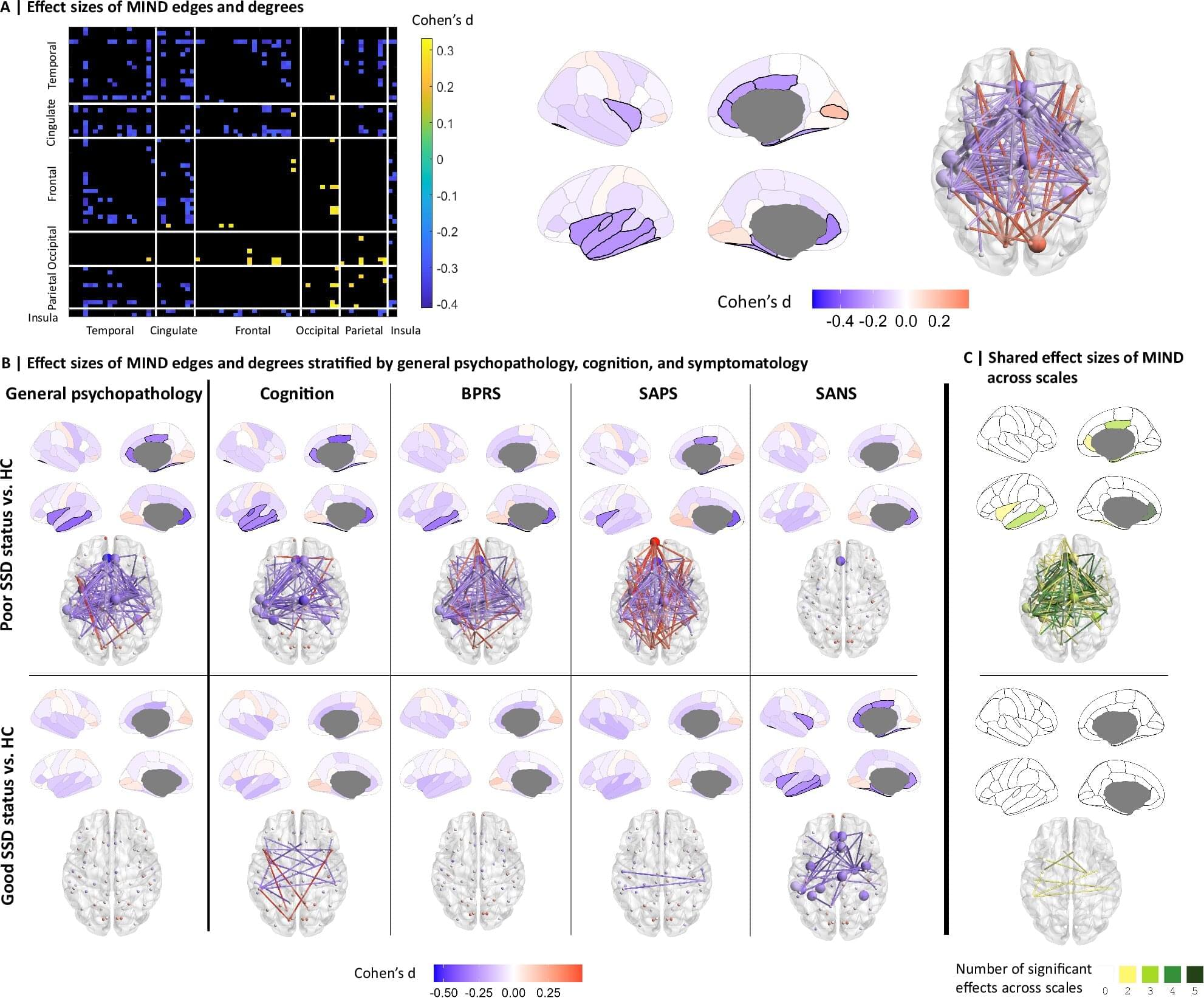
Researchers at the University of Seville have identified the possible origins of structural damage in the brains of patients with schizophrenia spectrum disorders (SSDs). These are regions that show the greatest morphological alterations in the early stages of the disease compared to neurotypical people of the same sex and age. The study also found that people with SSD have significant reductions in structural similarity between different regions of the temporal, cingulate and insular lobes.
The research is published in the journal Nature Communications.
The human brain is not a hard-wired machine but a malleable organ that is regularly re-shaping itself.
Neuroscientists at the University of Cambridge in the UK and the University of Pittsburgh in the US have now identified four major turning points in brain wiring between birth and death.
Like chapters of our lives, each of these neurological ‘epochs’ marks a new era of development or decline.