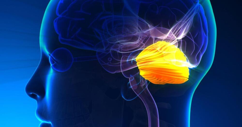
The cerebellum may coordinate thoughts much like it coordinates fluid movements.
What are biomarkers? They are medical signals that can measure health in an accurate and reproducible way. Common examples include blood pressure readings, heart rate, and even genetic test results.
Modern digital devices measure several health parameters. Fitbit trackers use sensors such as accelerometers to tell how many steps we’ve taken in a day or how fast we’ve been walking. When can such novel health measures function as medical biomarkers?
The measures must be objective, quantifiable, and reproducible. Additionally, scientific evidence needs to show that the health attribute measured by the device maps consistently and accurately to a clinical outcome. For example, voice signals from a smartphone’s microphone can detect mild cognitive impairment due to Alzheimer’s disease. World War II, commanders and troops communicated using hand-sent Morse codes. To avoid capture by enemies, telegraph operators had to remain anonymous. Any clues about operator identity or location could influence battle outcomes.
New research published in Human Brain Mapping provides evidence of a shared neural mechanism that underlies sleep disturbance and mental disorders in preadolescents. The findings indicate that sleep disturbance and mental health problems are both related to the connectivity between and within two important brain networks.
“I noticed the importance of sleep years ago when I read several papers about the immediate amyloid protein deposition in the brain after short-term sleep deprivation. Amyloid is neurotoxic waste in the brain and needs to be transported out by cerebrospinal fluid,” said study author Ze Wang, an associate professor of diagnostic radiology and nuclear medicine at the University of Maryland School of Medicine.
“But cerebrospinal fluid is basically static most of the time. The best time to have more cerebrospinal fluid and increased flow rate is at night when you lay down and fall asleep. It is this time that our cerebral blood flow reduces. Because our brain has a fixed size, the reduction of cerebral blood flow creates space for cerebrospinal fluid and the inhomogeneous change of blood flow creates power for cerebrospinal fluid to flow and then transport the neural waste out. This is why our brain generates two times as much cerebrospinal fluid at night than daytime.”
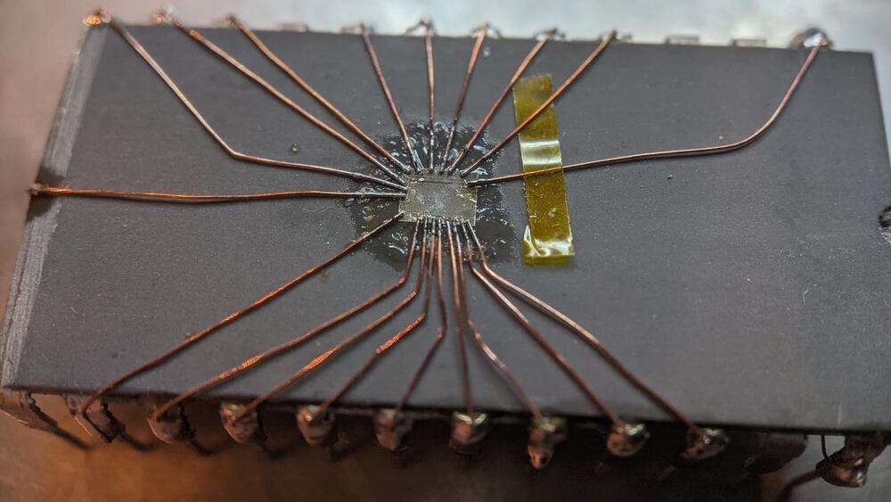
We might be amidst a chip shortage, but if you enjoy reverse-engineering, there’s never a shortage of intriguing old chips to dig into – and the 2513N 5×7 character ROM is one such chip. Amidst a long thread probing a few of these (Twitter, ThreadReader link), [TubeTime] has realized that two address lines were shorted inside of the package. A Twitter dopamine-fueled quest for truth has led them to try their hand at making the chip work anyway. Trying to clear the short with an external PSU led to a bond wire popping instead, as evidenced by the ESD diode connection disappearing.
A dozen minutes of sandpaper work resulted in the bare die exposed, making quick work of the bond wires as a side effect. Apparently, having the bond pads a bit too close has resulted in a factory defect where two of the pads merged together. No wonder the PSU wouldn’t take that on! Some X-acto work later, the short was cleared. But without the bond wires, how would [TubeTime] connect to it? This is where the work pictured comes in. Soldering to the remains of the bond wires has proven to be fruitful, reviving the chip enough to continue investigating, even if, it appears, it was never functional to begin with. The thread continued on with comparing ROMs from a few different chips [TubeTime] had on hand and inferences on what could’ve happened that led to this IC going out in the wild.
Such soldering experiments are always fun to try and pull off! We rarely see soldering on such a small scale, as thankfully, it’s not always needed, but it’s a joy to witness when someone does IC or PCB microsurgery to fix factory defects that render our devices inoperable before they were even shipped. Each time that a fellow hacker dares to grind the IC epoxy layers down and save a game console or an unidentified complex board, the world gets a little brighter. And if you aren’t forced to do it for repair reasons, you can always try it in an attempt to build the smallest NES in existence!

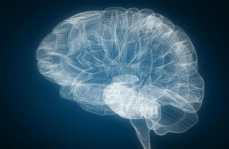
𝙎𝙡𝙚𝙚𝙥 𝙢𝙖𝙮 𝙗𝙚 𝙤𝙣𝙚 𝙤𝙛 𝙩𝙝𝙚 𝙢𝙤𝙨𝙩 𝙥𝙤𝙩𝙚𝙣𝙩 𝙢𝙚𝙙𝙞𝙘𝙞𝙣𝙚𝙨 𝙛𝙤𝙧 𝙩𝙝𝙚 𝙗𝙧𝙖𝙞𝙣, 𝙨𝙘𝙞𝙚𝙣𝙩𝙞𝙨𝙩𝙨 𝙖𝙧𝙚 𝙙𝙞𝙨𝙘𝙤𝙫𝙚𝙧𝙞𝙣𝙜, 𝙖𝙨 𝙩𝙝𝙚𝙮 𝙚𝙭𝙥𝙡𝙤𝙧𝙚 𝙩𝙝𝙚 𝙞𝙣𝙣𝙚𝙧 𝙡𝙖𝙗𝙮𝙧𝙞𝙣𝙩𝙝𝙨 𝙤𝙛 𝙩𝙝𝙚 𝙩𝙝𝙧𝙚𝙚-𝙥𝙤𝙪𝙣𝙙 𝙤𝙧𝙜𝙖𝙣 𝙙𝙪𝙧𝙞𝙣𝙜 𝙙𝙚𝙚𝙥 𝙨𝙡𝙚𝙚𝙥 𝙖𝙣𝙙 𝙙𝙧𝙚𝙖𝙢 𝙘𝙮𝙘𝙡𝙚𝙨 𝙞𝙣 𝙗𝙤𝙩𝙝 𝙝𝙚𝙖𝙡𝙩𝙝 𝙖𝙣𝙙 𝙙𝙞𝙨𝙚𝙖𝙨𝙚.
The Neuro-Network.
𝐂𝐚𝐧 𝐝𝐞𝐞𝐩 𝐬𝐥𝐞𝐞𝐩 𝐡𝐞𝐥𝐩 𝐝𝐞𝐯𝐚𝐬𝐭𝐚𝐭𝐢𝐧𝐠 𝐛𝐫𝐚𝐢𝐧 𝐝𝐢𝐬𝐨𝐫𝐝𝐞𝐫𝐬? 𝐒𝐜𝐢𝐞𝐧𝐭𝐢𝐬𝐭𝐬 𝐬𝐭𝐮𝐝𝐲𝐢𝐧𝐠 𝐏𝐚𝐫𝐤𝐢𝐧𝐬𝐨𝐧’𝐬 𝐰𝐚𝐧𝐭 𝐭𝐨 𝐟𝐢𝐧𝐝 𝐨𝐮𝐭
𝙈𝙚𝙙𝙞𝙘𝙖𝙓𝙥𝙧𝙚𝙨𝙨:
Sleep may be one of the most potent medicines for the brain, scientists are discovering, as they explore the inner labyrinths of the three-pound organ during deep sleep and dream cycles in both health and disease.
Watch the newest video from Big Think: https://bigth.ink/NewVideo.
Join Big Think Edge for exclusive videos: https://bigth.ink/Edge.
ABOUT BIG THINK:
Smarter Faster™
Big Think is the leading source of expert-driven, actionable, educational content — with thousands of videos, featuring experts ranging from Bill Clinton to Bill Nye, we help you get smarter, faster. Subscribe to learn from top minds like these daily. Get actionable lessons from the world’s greatest thinkers & doers. Our experts are either disrupting or leading their respective fields. We aim to help you explore the big ideas and core skills that define knowledge in the 21st century, so you can apply them to the questions and challenges in your own life.
Other Frequent contributors include Michio Kaku & Neil DeGrasse Tyson.
Michio Kaku Playlist: https://bigth.ink/kaku.
Bill Nye Playlist: https://bigth.ink/BillNye.
Neil DeGrasse Tyson Playlist: https://bigth.ink/deGrasseTyson.
Join Big Think Edge, to gain access to a world-class learning platform focused on building the soft skills essential to 21st century success. It features insight from many of the most celebrated and intelligent individuals in the world today. Topics on the platform are focused on: emotional intelligence, digital fluency, health and wellness, critical thinking, creativity, communication, career development, lifelong learning, management, problem solving & self-motivation.
If you are a scientist, willing to share your science with curious teens, consider joining Lecturers Without Borders!
Established by three scientists, Luibov Tupikina, Athanasia Nikolau, and Clara Delphin Zemp, and high school teacher Mikhail Khotyakov, Lecturers Without Borders (LeWiBo) is an international volunteer grassroots organization that brings together enthusiastic science researchers and science-minded teens. LeWiBo founders noticed that scientists tend to travel a lot – for fieldwork, conferences, or lecturing – and realized scientists could be a great source of knowledge and inspiration to local schools. To this end, they asked scientists to volunteer for talks and workshops. The first lecture, delivered in Nepal in 2017 by two researchers, a mathematician and a climatologist, was a great success. In the next couple of years, LeWiBo volunteers presented at schools in Russia and Belarus; Indonesia and Uganda; India and Nepal. Then, the pandemic forced everything into the digital realm, bringing together scientists and schools across the globe. I met with two of LeWiBo’s co-founders, physicist Athanasia Nikolaou and math teacher Mikhail Khotyakov, as well as their coordinator, Anastasia Mityagina, to talk about their offerings and future plans.
Julia Brodsky: So, how many people volunteer for LeWiBo at this time?
Anastasia Mityagina: We have over 200 scientists in our database. This year alone, volunteers from India, Mozambique, Argentina, the United States, France, Egypt, Israel, Brazil, Ghana, Nigeria, Ethiopia, Botswana, Portugal, Croatia, Malaysia, Spain, Colombia, Italy, Germany, Greece, Denmark, Poland, the United Kingdom, Austria, Albania, Iran, Mexico, Russia, and Serbia joined us. Their areas of expertise vary widely, from informatics, education, and entrepreneurship, to physics, chemistry, space and planetary sciences, biotechnology, oceanography, viral ecology, water treatment, nanotechnology, artificial intelligence, astrobiology, neuroscience, and sustainability. We collaborate with hundreds of schools, education centers, and science camps for children in different parts of the world. In addition, our network includes more than 50 educational associations in 48 countries that help us reach out to approximately 8,000 schools worldwide.
The microbots are applied nasally to treat brain diseases.
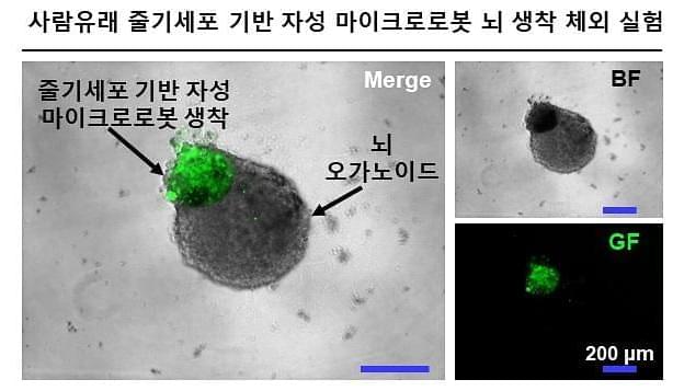
Scientists have successfully guided a microbot through the nasal pathways to the brain of a mouse. If the same approach can be replicated in humans, it could be a game-changer against neurodegenerative disease, enabling doctors to deliver therapies directly to the brain.
A research team led by DGIST (the Daegu Gyeongbuk Institute of Science and Technology in South Korea) has created a microrobot propelled by magnets that can navigate the human body. The trial, published in the journal Advanced Materials, describes how they manufactured the microrobot, dubbed a Cellbot, by magnetizing stem cells extracted from the human nasal cavity. The scientists then tested the ability of the Cellbot to move through the body’s confined vessels and passages to reach its target, which it completed with ease.
DGIST said in a statement that “This approach has the potential to effectively treat central nervous system disorders in a minimally invasive manner.”