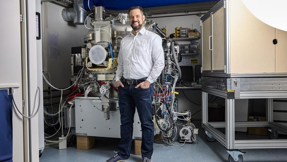
A place for cutting-edge research: PSI researchers to receive comprehensive funding from the US NIH for their brain research.


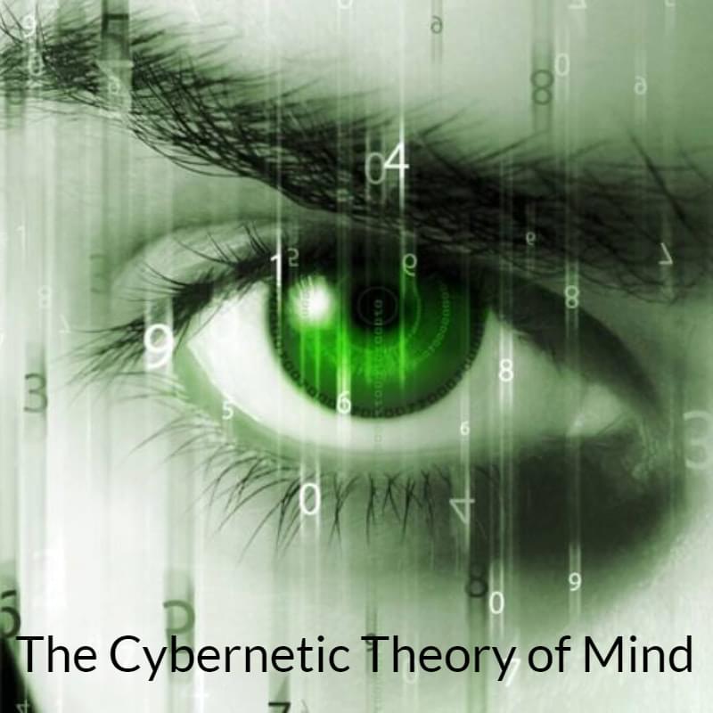
It wouldn’t shock me if all the buzz around searching for the ‘locus of consciousness’ merely fine-tunes our grasp of how the brain is linked to consciousness — without actually revealing where consciousness comes from, because it’s not generated in the brain. Similarly, your smartphone doesn’t create the Internet or a cellular network; it just processes them. Networks of minds are a common occurrence throughout the natural world. What sets humans apart is the impending advent of cybernetic connectivity explosion that could soon evolve into a form of synthetic telepathy, eventually leading to the rise of a unified, global consciousness — what could be termed the Syntellect Emergence.
#consciousness #phenomenology #cybernetics #cognition #neuroscience
In summary, the study of consciousness could be conceptualized through a variety of lenses: as a series of digital perceptual snapshots, as a cybernetic system with its feedback processes, as a grand theater; or perhaps even as a VIP section in a cosmological establishment of magnificent complexity. Today’s leading theories of consciousness are largely complementary, not mutually exclusive. These multiple perspectives not only contribute to philosophical discourse but also herald the dawn of new exploratory avenues, equally enthralling and challenging, in our understanding of consciousness.
In The Cybernetic Theory of Mind (2022), I expand on existing theories to propose certain conceptual models and concepts, such as Noocentrism, Digital Presentism (D-Theory of Time), Experiential Realism, Ontological Holism, Multi-Ego Pantheistic Solipsism, the Omega Singularity, deeming a non-local consciousness, or Universal Mind, as the substrate of objective reality. In search of God’s equation, we finally look upward for the source. What many religions call “God” is clearly an interdimensional being within the nested levels of complexity. Besides setting initial conditions for our universe, God speaks to us in the language of religion, spirituality, synchronicities and transcendental experiences.
Summary: Neuroscientists delved into the mechanisms behind true and false memories. Their study reveals that electrical signals in the hippocampus can differentiate between the imminent recall of authentic versus fabricated memories.
By monitoring neural activity in epilepsy patients, the team identified distinct patterns ahead of a correct or false recall. These findings not only offer insights into memory retrieval but may also pave the way for novel therapeutic interventions for disorders like PTSD.

The question comes down to this: If materialism collapses, what will science look like? Will the people who are interested in science today continue to be so? Will the same people continue to dominate?
One thing for sure: A lot of things will come tumbling out in the wash.
*In my experience, the abortion issue has mostly been Catholic and other grannies vs. abortionists. If, like David Chalmers, you are inclined to take bets, bet on the grannies.
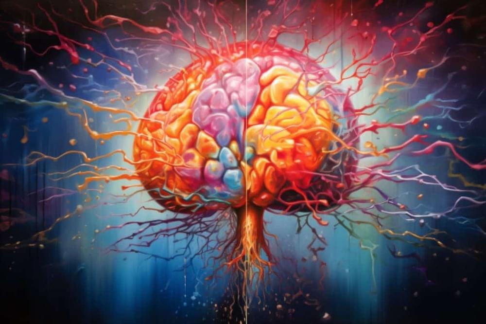
The human brain can change – but usually only slowly and with great effort, such as when learning a new sport or foreign language, or recovering from a stroke. Learning new skills correlates with changes in the brain, as evidenced by neuroscience research with animals and functional brain scans in people. Presumably, if you master Calculus 1, something is now different in your brain. Furthermore, motor neurons in the brain expand and contract depending on how often they are exercised – a neuronal reflection of “use it or lose it.”
People may wish their brains could change faster – not just when learning new skills, but also when overcoming problems like anxiety, depression and addictions.
… More
Change in the brain usually comes with plenty of effort over time. Neuroscientists are working to understand how psychedelic drugs provide a shortcut that seems to rely on existing brain systems.
Participants individually interacted with a conversational AI mental health chatbot for about 30 minutes to determine if they would recommend it to a friend.
As human beings, we rely on recommendations or warnings from our friends and family. It gives us an added perspective on what to expect from a particular service, a product, or another human being. As per the latest study, the same is true for the way in which we trust and perceive an AI chatbot.
Researchers from Massachusetts Institute of Technology (MIT) and Arizona State University conducted a study in which they found that even though every person in their sample size of 310 people interacted with the exact same chatbot, their interactions with it were influenced by what they had been told before.
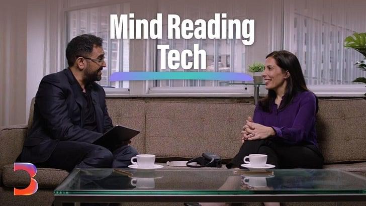
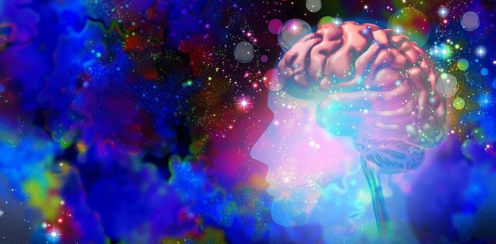
“New devices can read and manipulate our mental states to help us relax, learn and reduce pain. As they do this, they harvest data. Can businesses be trusted with this private information? How can we make use of this technology while protecting the last fortress of our humanity — our thoughts and emotions?”
As neurotechnology becomes widely accessible, do we need to legally protect our thoughts?