Summary: A comprehensive study mapped neuronal IL-1R1 (nIL-1R1) expression in the mouse brain, highlighting its role in sensory processing, mood, and memory regulation. Researchers found that neurons expressing IL-1R1 integrate immune and neural signals, revealing connections between inflammation and brain disorders like depression and anxiety.
The study pinpointed key regions, such as the somatosensory cortex and hippocampus, where IL-1 signaling influences synapse organization and neural circuit modulation. Notably, neuronal IL-1R1 modifies synaptic pathways without triggering inflammation, suggesting distinct functions in the central nervous system.
I want to look at what Dennett has to say about patterns because 1) I introduced the term in my previous discussion, In Search of Dennett’s Free-Floating Rationales [1], and 2) it is interesting for what it says about his philosophy generally.
You’ll recall that, in that earlier discussion, I pointed out talk of “free-floating rationales” (FFRs) was authorized by the presence of a certain state of affairs, a certain pattern of relationships among, in Dennett’s particular example, an adult bird, (vulnerable) chicks, and a predator. Does postulating talk of FFRs add anything to the pattern? Does it make anything more predictable? No. Those FFRs are entirely redundant upon the pattern that authorizes them. By Occam’s Razor, they’re unnecessary.
With that, let’s take a quick look at Dennett’s treatment of the role of patterns in his philosophy. First I quote some passages from Dennett, with a bit of commentary, and then I make a few remarks on my somewhat different treatment of patterns. In a third post I’ll be talking about the computational capacities of the mind/brain.
As an embryo grows, there is a continuous stream of communication between cells to form tissues and organs. Cells need to read numerous cues from their environment, and these may be chemical or mechanical in nature. However, these alone cannot explain collective cell migration, and a large body of evidence suggests that movement may also happen in response to embryonic electrical fields. How and where these fields are established within embryos was unclear until now.
“We have characterized an endogenous bioelectric current pattern, which resembles an electric field during development, and demonstrated that this current can guide migration of a cell population known as the neural crest,” highlights Dr. Elias H. Barriga, the corresponding author who led the study published in Nature Materials.
Initially, Dr. Barriga and his team began research on the neural crest at the former Gulbenkian Institute of Science (IGC) in Oeiras, Portugal before continuing research in Dresden, establishing a group at the Cluster of Excellence Physics of Life.
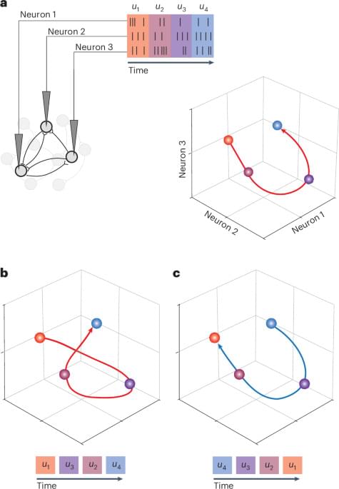
Oby, Degenhart, Grigsby and colleagues used a brain–computer interface to challenge monkeys to override their natural time courses of neural activity. They found the time courses to be highly robust, suggestive of network-level computational mechanisms.
Large language models surpass human experts in predicting neuroscience results, according to a study published in Nature Human Behaviour.
Scientific research is increasingly challenging due to the immense growth in published literature. Integrating noisy and voluminous findings to predict outcomes often exceeds human capacity. This investigation was motivated by the growing role of artificial intelligence in tasks such as protein folding and drug discovery, raising the question of whether LLMs could similarly enhance fields like neuroscience.
Xiaoliang Luo and colleagues developed BrainBench, a benchmark designed to test whether LLMs could predict the results of neuroscience studies more accurately than human experts. BrainBench included 200 test cases based on neuroscience research abstracts. Each test case consisted of two versions of the same abstract: one was the original, and the other had a modified result that changed the study’s conclusion but kept the rest of the abstract coherent. Participants—both LLMs and human experts—were tasked with identifying which version was correct.
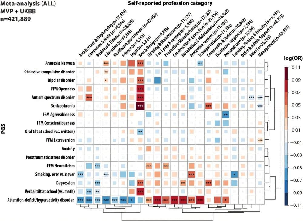
Polygenic scores (PGS) are metrics used to estimate the genetic predisposition of people to developing specific mental health conditions, personality traits or diseases. In recent years, these metrics have often been used to investigate the intricate connections between genes and environmental factors.
Researchers at the JJ Peters VA Medical Center, Icahn School of Medicine at Mount Sinai and other institutes recently carried out a study aimed at determining whether neuropsychiatric polygenic scores could predict the professional categories that individuals belong to. Their findings, published in Nature Human Behaviour, suggest that these scores weakly predict the professional category that people belong to.
“Neuropsychiatric disorders are both common and highly heritable, yet they remain heavily stigmatized,” Georgios Voloudakis, first author of the paper, told Medical Xpress.
Leading A Government-Wide Response To Long COVID — Dr. Ian Simon, Ph.D. — Director, Office of Long COVID Research and Practice, Office of the Assistant Secretary for Health (OASH), U.S. Department of Health and Human Services (HHS)
Dr. Ian Simon, Ph.D. is the Director for the Office of Long COVID Research and Practice (https://www.hhs.gov/longcovid/index.html), in the Office of Science and Medicine, in the Office of the Assistant Secretary for Health at the U.S. Department of Health \& Human Services.
The Office of Science and Medicine harnesses the power of collaboration, scientific analysis, data-driven innovation, and emerging technologies for advancing initiatives across the Department, including not just Long COVID, but in the areas of behavioral health, health equity, kidney disease, infection-associated chronic conditions, mother-infant dyad, sickle cell disease, and traumatic brain injury.
Previously Dr. Simon was the Assistant Director for Health Strategy and Biopreparedness at the White House Office of Science and Technology Policy, where he led pandemic prevention and biosecurity policy priorities. Most recently, he was the Senior Advisor to the Director of NIH’s National Institute of Allergy and Infectious Diseases (NIAID).
Prior to working at NIAID, Dr. Simon was the Assistant Director of the Institute for Defense Analyses (IDA) Science and Technology Policy Institute. In that role, he specialized in developing policy initiatives including bioeconomy, STEM education, pandemic preparedness, biosecurity, and international S\&T cooperation.
Stimulating dopamine-producing brain cells wirelessly with gold nanoparticles has proven effective at treating mice with Parkinson’s disease, even reversing a portion of their neurological damage.
Researchers from the National Center for Nanoscience and Technology of China (NCNST) say it’s a significant step forward for using brain simulation to tackle Parkinson’s in humans, a neurodegenerative condition that affects more than 10 million people worldwide.
Deep inside the brains of those with the condition, dopamine-producing neurons take a major hit as insoluable clumps of a protein called alpha-synuclein accumulate, gradually depriving patients of an ability to control their movements.
Glioblastoma, an aggressive and often fatal form of brain cancer, has long posed a formidable challenge to doctors and patients alike. Yet, a groundbreaking clinical trial is offering a glimmer of hope, capturing global attention for its potential to revolutionize cancer treatment. A 62-year-old engineer, faced with a grim prognosis, has experienced something extraordinary—his tumour has shrunk significantly in a matter of weeks. This remarkable outcome marks the beginning of a journey that could redefine how we treat one of the most challenging cancers. What makes this approach so promising, and how could it change the future for patients?
Glioblastoma, often referred to as glioblastoma multiforme (GBM), is the most aggressive and common form of primary brain cancer in adults. Originating from glial cells—specifically astrocytes that support nerve cells—this malignancy is notorious for its rapid growth and diffuse infiltration into surrounding brain tissue, making complete surgical removal challenging.
While research continues on the potential of psychedelics as a clinical treatment, a recent study highlights the need to better understand their adverse effects.
PsychiatryOnline subscription options offer access to the DSM-5-TR® library, books, journals, CME, and patient resources. This all-in-one virtual library provides psychiatrists and mental health professionals with key resources for diagnosis, treatment, research, and professional development.
Need more help? PsychiatryOnline Customer Service may be reached by emailing [email protected] or by calling 800−368−5777 (in the U.S.) or 703−907−7322 (outside the U.S.).