Toggle light / dark theme

Will AI make us crazy?

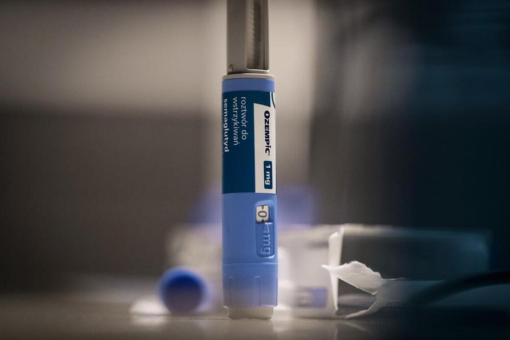
Coverage of the risks and benefits of AI have paid scant attention to how chatbots might affect public health at a time when depression, suicide, anxiety, and mental illness are epidemic in the United States. But mental health experts and the healthcare industry view AI mostly as a promising tool, rather than a potential threat to mental health.

Beyond Sight: Unraveling the Mysteries of Brain Wiring

Summary: Scientists made a novel discovery using zebrafish with a genetic mutation. These ‘deep-blind’ fish lack connections between the retina and brain yet retain functional brain circuits.

Remarkably, despite their inability to see, direct brain stimulation through optogenetics triggers normal visual behavior. This suggests that much of the zebrafish brain’s wiring is innate and doesn’t rely heavily on visual experience.

New method tracks how brain cells age

Hospital nurseries routinely place soft bands around the tiny wrists of newborns that hold important identifying information such as name, sex, mother, and birth date. Researchers at Rockefeller University are taking the same approach with newborn brain cells—but these neonates will keep their ID tags for life, so that scientists can track how they grow and mature, as a means for better understanding the brain’s aging process.

As described in a new paper in Cell, the new method developed by Rockefeller geneticist Junyue Cao and his colleagues is called TrackerSci (pronounced “sky”). This low-cost, high-throughput approach has already revealed that while newborn cells continue to be produced through life, the kinds of cells being produced greatly vary in different ages. This groundbreaking work, led by co-first authors Ziyu Lu and Melissa Zhang from Cao’s lab, promises to influence not only the study of the brain but also broader aspects of aging and disease across the human body.

“The cell is the basic functional unit of our body, so changes to the cell essentially underlie virtually every disease and the aging process,” says Cao, head of the Laboratory of Single-Cell Genomics and Population Dynamics. “If we can systematically characterize the different cells and their dynamics using this novel technique, we may get a panoramic view of the mechanisms of many diseases and the enigma of aging.”

Reactivation of Early-Life Stress-Sensitive Neuronal Ensembles Contributes to Lifelong Stress Hypersensitivity

Early-life stress (ELS) is one of the strongest lifetime risk factors for depression, anxiety, suicide, and other psychiatric disorders, particularly after facing additional stressful events later in life. Human and animal studies demonstrate that ELS sensitizes individuals to subsequent stress. However, the neurobiological basis of such stress sensitization remains largely unexplored. We hypothesized that ELS-induced stress sensitization would be detectable at the level of neuronal ensembles, such that cells activated by ELS would be more reactive to adult stress. To test this, we leveraged transgenic mice to genetically tag, track, and manipulate experience-activated neurons. We found that in both male and female mice, ELS-activated neurons within the nucleus accumbens (NAc), and to a lesser extent the medial prefrontal cortex, were preferentially reactivated by adult stress. To test whether reactivation of ELS-activated ensembles in the NAc contributes to stress hypersensitivity, we expressed hM4Dis receptor in control or ELS-activated neurons of pups and chemogenetically inhibited their activity during experience of adult stress. Inhibition of ELS-activated NAc neurons, but not control-tagged neurons, ameliorated social avoidance behavior following chronic social defeat stress in males. These data provide evidence that ELS-induced stress hypersensitivity is encoded at the level of corticolimbic neuronal ensembles.

SIGNIFICANCE STATEMENT Early-life stress enhances sensitivity to stress later in life, yet the mechanisms of such stress sensitization are largely unknown. Here, we show that neuronal ensembles in corticolimbic brain regions remain hypersensitive to stress across the life span, and quieting these ensembles during experience of adult stress rescues stress hypersensitivity.

Brain implants, software guide speech-disabled person’s intended words to computer screen

Pat Bennett’s prescription is a bit more complicated than “Take a couple of aspirins and call me in the morning.” But a quartet of baby-aspirin-sized sensors implanted in her brain are aimed at addressing a condition that’s frustrated her and others: the loss of the ability to speak intelligibly. The devices transmit signals from a couple of speech-related regions in Bennett’s brain to state-of-the-art software that decodes her brain activity and converts it to text displayed on a computer screen.

Bennett, now 68, is a former human resources director and onetime equestrian who jogged daily. In 2012, she was diagnosed with amyotrophic lateral sclerosis, a progressive neurodegenerative disease that attacks neurons controlling movement, causing physical weakness and eventual paralysis.


Our brains remember how to formulate words even if the muscles responsible for saying them out loud are incapacitated. A brain-computer hookup is making the dream of restoring speech a reality.

Scientists Unveil Unprecedented “Live” View Into the Brain’s Complexity

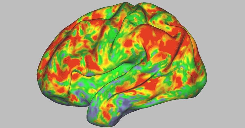
The human brain, with its intricate network of approximately 86 billion neurons, is arguably among the most complex specimens scientists have ever encountered. It holds an immense, yet currently immeasurable, wealth of information, positioning it as the pinnacle of computational devices.

Grasping this level of intricacy is challenging, making it essential for us to employ advanced technologies that can decode the minute, intricate interactions happening within the brain at microscopic levels. Thus, imaging emerges as a pivotal instrument in the realm of neuroscience.

The new imaging and virtual reconstruction technology developed by Johann Danzl’s group at ISTA is a big leap in imaging brain activity and is aptly named LIONESS – Live Information Optimized Nanoscopy Enabling Saturated Segmentation. LIONESS is a pipeline to image, reconstruct, and analyze live brain tissue with a comprehensiveness and spatial resolution not possible until now.

Science Fiction Meets Neuro-Reality: Organoids to Rebuild the Brain

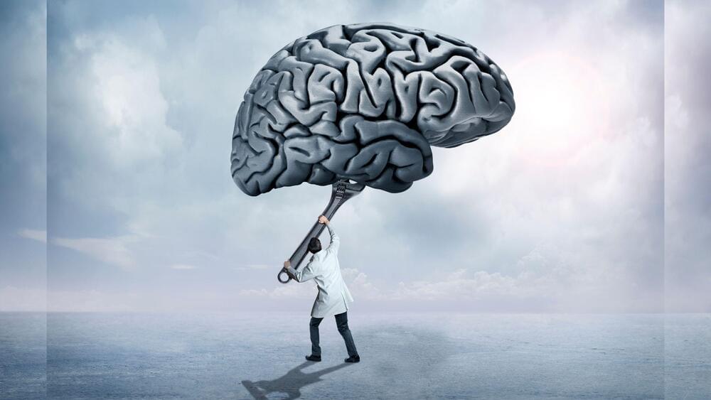
This is leading to even better brain engineering 👏 🙌 👌 😀 😄.


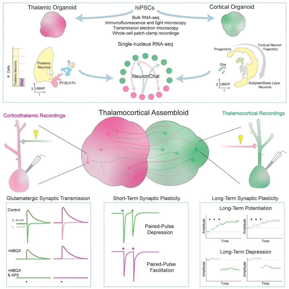
Computer-augmented brains, cures to blindness, and rebuilding the brain after injury all sound like science fiction. Today, these disruptive technologies aren’t just for Netflix, “Terminator,” and comic book fodder — in recent years, these advances are closer to reality than some might realize, and they have the ability to revolutionize neurological care.

Neurologic disease is now the world’s leading cause of disability, and upwards of 11 million people have some form of permanent neurological problem from traumatic brain injuries and stroke. For example, if a traumatic brain injury has damaged the motor cortex — the region of the brain involved in voluntary movements — patients could become paralyzed, without hope of regaining full function. Or some stroke patients can suffer from aphasia, the inability to speak or understand language, due to damage to the brain regions that control speech and language comprehension.

Thanks to recent advances, sometimes lasting neurologic disease can be prevented. For example, if a stroke patient is seen quickly enough, life-threatening or-altering damage can be avoided, but it’s not always possible. Current treatments to most neurologic disease are fairly limited, as most therapies, including medications, aim to improve symptoms but can’t completely recover lost brain function.

/* */