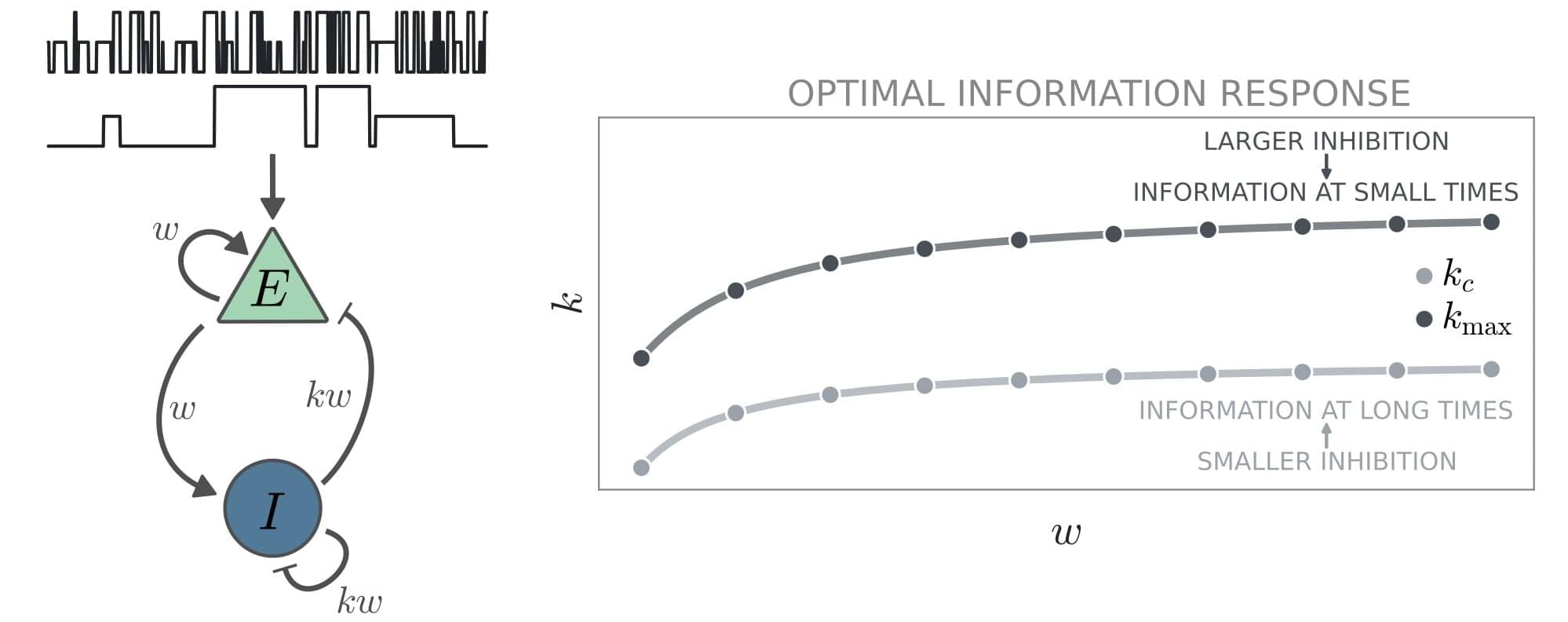
Toggle light / dark theme

As long as people have been alive, they’ve wanted to stay alive. But unlike finding the fountain of youth or becoming a vampire, uploading your brain to a computer or the cloud might actually be possible. Theoretically, we already know how to do it, and Elon Musk is even trying a brain implant with Neuralink. But technically, we have a long way to go. We explain the main technological advancements that we’ll need to make whole brain emulation a reality.

MORE TECHNOLOGY VIDEOS:
Why We Still Don’t Have Smart Contact Lenses.


Why We Still Haven’t Cloned Humans — It’s Not Just Ethics.

How Blockchain And Remote Monitoring Can Improve The Healthcare Experience.

#Brain #Tech #TechInsider.

Tech Insider tells you all you need to know about tech: gadgets, how-to’s, gaming, science, digital culture, and more.

Visit us at: https://www.businessinsider.com.
TI on Facebook: https://www.facebook.com/techinsider.
TI on Instagram: https://www.instagram.com/tech_insider/
TI on Twitter: https://twitter.com/techinsider.
TI on Amazon Prime: http://read.bi/PrimeVideo.
INSIDER on Snapchat: https://insder.co/2KJLtVo.

What it’ll take to upload our brains to a computer.

Until now, it was unclear as to whether drugs that clear amyloid, which accumulate in the brain during aging and accompany diseases such as Alzheimer’s, have any influence over cognitive decline.

Previous studies have aimed to find this out, but results have been inconclusive due to study designs, hard-to-interpret data, and other issues that muddy the waters. March 10-14th saw the 15th International Conference on Alzheimer’s and Parkinson’s Diseases being held (virtually of course), where Dr. Mark Mintun of Eli Lilly presented data that, at least somewhat, affirmatively answers the question [1].

Alzheimer’s disease is a neurodegenerative disease that affects millions of people worldwide. It is characterized by the accumulation of amyloid plaques and disordered protein fibers called tau tangles in the brain, which lead to cognitive impairment and dementia. Scientists have long been trying to understand the underlying mechanisms behind Alzheimer’s disease and find effective treatments for the condition.

This video is my take on 3B1B’s Summer of Math Exposition (SoME) competition.

It explains in pretty intuitive terms how ideas from topology (or “rubber geometry”) can be used in neuroscience, to help us understand the way information is embedded in high-dimensional representations inside neural circuits.

OUTLINE:
00:00 Introduction.
01:34 — Brief neuroscience background.
06:23 — Topology and the notion of a manifold.
11:48 — Dimension of a manifold.
15:06 — Number of holes (genus)
18:49 — Putting it all together.

____________
Main paper:
Chaudhuri, R., Gerçek, B., Pandey, B., Peyrache, A. & Fiete, I. The intrinsic attractor manifold and population dynamics of a canonical cognitive circuit across waking and sleep. Nat Neurosci 22, 1512–1520 (2019).

_________________________
Other relevant references:
1. Jazayeri, M. & Ostojic, S. Interpreting neural computations by examining intrinsic and embedding dimensionality of neural activity. arXiv:2107.04084 [q-bio] (2021).
2. Gallego, J. A., Perich, M. G., Chowdhury, R. H., Solla, S. A. & Miller, L. E. Long-term stability of cortical population dynamics underlying consistent behavior. Nat Neurosci 23260–270 (2020).
3. Bernardi, S. et al. The Geometry of Abstraction in the Hippocampus and Prefrontal Cortex. Cell 183954–967.e21 (2020).
4. Shine, J. M. et al. Human cognition involves the dynamic integration of neural activity and neuromodulatory systems. Nat Neurosci 22289–296 (2019).
5. Remington, E. D., Narain, D., Hosseini, E. A. & Jazayeri, M. Flexible Sensorimotor Computations through Rapid Reconfiguration of Cortical Dynamics. Neuron 98, 1005–1019.e5 (2018).
6. Low, R. J., Lewallen, S., Aronov, D., Nevers, R. & Tank, D. W. Probing variability in a cognitive map using manifold inference from neural dynamics. http://biorxiv.org/lookup/doi/10.1101/418939 (2018) doi:10.1101/418939.
7. Elsayed, G. F., Lara, A. H., Kaufman, M. T., Churchland, M. M. & Cunningham, J. P. Reorganization between preparatory and movement population responses in motor cortex. Nat Commun 7, 13239 (2016).
8. Peyrache, A., Lacroix, M. M., Petersen, P. C. & Buzsáki, G. Internally organized mechanisms of the head direction sense. Nat Neurosci 18569–575 (2015).
9. Dabaghian, Y., Mémoli, F., Frank, L. & Carlsson, G. A Topological Paradigm for Hippocampal Spatial Map Formation Using Persistent Homology. PLoS Comput Biol 8, e1002581 (2012).
10. Yu, B. M. et al. Gaussian-Process Factor Analysis for Low-Dimensional Single-Trial Analysis of Neural Population Activity. Journal of Neurophysiology 102614–635 (2009).
11. Singh, G. et al. Topological analysis of population activity in visual cortex. Journal of Vision 8, 11–11 (2008).

The majority of animations in this video were made using Manim — an open source python library (github.com/ManimCommunity/manim) and brainrender (github.com/brainglobe/brainrender)

Join us on Patreon! https://www.patreon.com/MichaelLustgartenPhD

Discount Links:
NAD+ Quantification: https://www.jinfiniti.com/intracellular-nad-test/
Use Code: ConquerAging At Checkout.

Green Tea: https://www.ochaandco.com/?ref=conqueraging.

Oral Microbiome: https://www.bristlehealth.com/?ref=michaellustgarten.

Epigenetic Testing: https://bit.ly/3Rken0n.
Use Code: CONQUERAGING!

At-Home Blood Testing: https://getquantify.io/mlustgarten.

In a recent study published in the journal Maturitas, researchers conducted a systematic review and meta-analysis to compare the risk of cardiovascular events in women with and without endometriosis.

Study: Endometriosis and cardiovascular disease: A systematic review and meta-analysis. Image Credit: Bangkok Click Studio / Shutterstock.

Apart from autoimmune disorders, polycystic ovary syndrome, depression, and premature menopause, there are pregnancy-associated risk factors for cardiovascular diseases, such as gestational diabetes, pregnancy-related hypertensive disorders, placental abruption, preterm delivery, and pregnancy loss. Women experience a higher mortality rate due to cardiovascular diseases, and while the treatment methods are the same for men and women, the presentation, symptoms, diagnosis, risk factors, and response to treatment differ for women.

How can flesh and blood brains give rise to pains and pleasures, dreams and desires, sights and sounds? Some believe this ‘hard problem’ of consciousness can never be solved. Can we expect any breakthroughs as the science of the mind progresses?

Our annual debate this year considers whether the problem of consciousness really is intractable. Our illustrious panel is neuroscientist Anil Seth and philosophers Louise Antony, Maja Spener and Philip Goff, with the BBC’s Ritula Shah chairing.

Speakers.
Anil Seth is Professor of Cognitive and Computational Neuroscience at the University of Sussex.
Louise Antony is Professor Emerita at the University of Massachusetts, Amherst.
Maja Spener is Associate Professor in Philosophy at the University of Birmingham.
Philip Goff is Associate Professor in the Department of Philosophy at Durham University.

Chair.
Ritula Shah is a journalist and presenter of The World Tonight on BBC Radio 4.

Synaptic plasticity is a critical process that regulates neuronal activity by allowing neurons to adjust their synaptic strength in response to changes in activity. Despite the high proximity of excitatory glutamatergic and inhibitory GABAergic postsynaptic zones and their functional integration within dendritic regions, concurrent plasticity has historically been underassessed. Growing evidence for pathological disruptions in the excitation and inhibition (E/I) balance in neurological and neurodevelopmental disorders indicates the need for an improved, more “holistic” understanding of synaptic interplay. There continues to be a long-standing focus on the persistent strengthening of excitation (excitatory long-term potentiation; eLTP) and its role in learning and memory, although the importance of inhibitory long-term potentiation (iLTP) and depression (iLTD) has become increasingly apparent. Emerging evidence further points to a dynamic dialogue between excitatory and inhibitory synapses, but much remains to be understood regarding the mechanisms and extent of this exchange. In this mini-review, we explore the role calcium signaling and synaptic crosstalk play in regulating postsynaptic plasticity and neuronal excitability. We examine current knowledge on GABAergic and glutamatergic synapse responses to perturbances in activity, with a focus on postsynaptic plasticity induced by short-term pharmacological treatments which act to either enhance or reduce neuronal excitability via ionotropic receptor regulation in neuronal culture. To delve deeper into potential mechanisms of synaptic crosstalk, we discuss the influence of synaptic activity on key regulatory proteins, including kinases, phosphatases, and synaptic structural/scaffolding proteins. Finally, we briefly suggest avenues for future research to better understand the crosstalk between glutamatergic and GABAergic synapses.

Ligand-gated ion channel GABA type A receptors (GABAARs) mediate the majority of fast inhibition in the central nervous system, while glutamatergic AMPA receptors (AMPARs) and NMDA receptors (NMDARs) collectively mediate fast excitatory neurotransmission. NMDARs particularly play a unique role in synaptic plasticity due to high calcium permeability and voltage-dependent Mg2+ block typically relieved by AMPAR-mediated depolarization. Slow inhibition and excitation are generated by G protein-coupled, GABA type B (GABABRs) and metabotropic glutamate receptors (mGluRs), respectively. The concerted action of these receptors balances neuronal excitability. A close and coordinated spatial relationship between glutamatergic and GABAergic synapses on dendrites (Megías et al., 2001; Bleckert et al., 2013; Iascone et al., 2020), sometimes as near as on the same spine (Chen et al., 2012), facilitates synaptic input integration, dynamic calcium regulation, synaptic crosstalk, and coregulation.

Synaptic plasticity describes the ability of synapses to adapt their relative strength based on the overall level of activity or specific activity patterns, often by dynamic regulation of receptor-synaptic scaffold interactions or through trafficking. During development, it is heavily involved in dendritic growth, synaptogenesis, and the formation of neural circuits (reviewed in Akgül and McBain, 2016; Ismail et al., 2017; Jenks et al., 2021). In mature neurons, synaptic plasticity is responsible for synapse remodeling during experience. Genetic mutations or pathology leading to altered excitatory or inhibitory neurotransmission or impaired synaptogenesis typically result in deficits in synaptic plasticity, a common feature in neurodevelopmental and neurological disorders (Rudolph and Möhler, 2014; Mele et al., 2019), including autism (Hansel, 2019; Sohal and Rubenstein, 2019), down syndrome (Galdzicki et al., 2001; Schulz et al.