American composer Alvin Lucier was well-known for his experimental works that tested the boundaries of music and art. A longtime professor at Wesleyan University (before retiring in 2011), Alvin passed away in 2021 at the age of 90. However, that wasn’t the end of his lifelong musical odyssey.
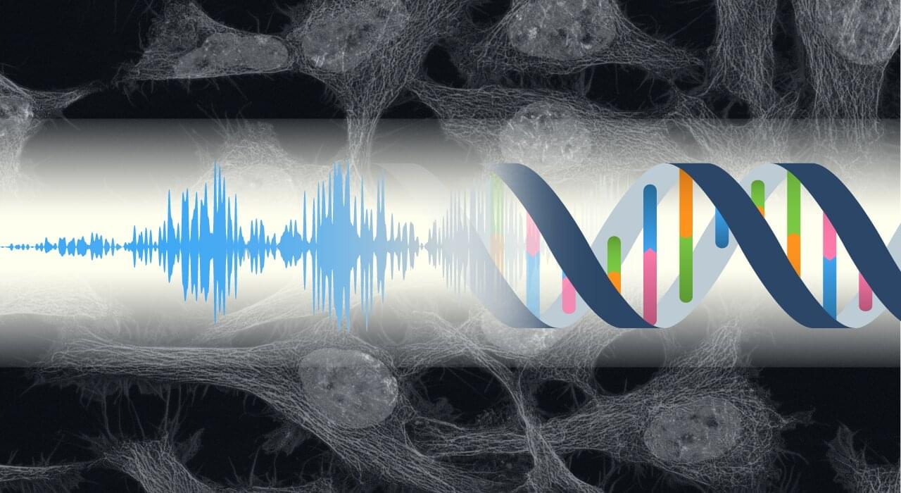
Earlier this month, at the Art Gallery of Western Australia, a new art installation titled Revivification used Lucier’s “brain matter”—hooked up to an electrode mesh connected to twenty large brass plates—to create electrical signals that triggered a mallet to strike the varying plates, creating a kind of post-mortem musical piece. Conceptualized in collaboration with Lucier himself before his death, the artists solicited the help of researchers from Harvard Medical School, who grew a mini-brain from Lucier’s white blood cells. The team created stem cells from these white blood cells, and due to their pluripotency, the cells developed into cerebral organoids somewhat similar to developing human brains.