One of the co-founders of Elon Musk’s Neuralink Corp. is building a different kind of brain implant.
Category: neuroscience – Page 380

SERTM2: a neuroactive player in the world of micropeptides
We characterized the 3’ end sequence of the lncMN3 transcript using rapid amplification of cDNA ends (RACE) and we confirmed the existence of the annotated isoform (Appendix Fig. S1A).
The analysis of the expression profile of lncMN3 during mESCs in vitro differentiation to MNs indicated that while it is not expressed in mESCs, it starts to be present in embryoid bodies at day 5 (EB5), reaches its maximum in EBs at day 6 (EB6) and decreases in the mixed population containing MNs (DIV3) obtained after cells dissociation (Fig. 1B). The observed decrease in expression is probably caused by a dilution effect due to the experimental protocol used for MN differentiation (Wichterle and Peljto, 2008) rather than to a real down-regulation. In fact, the MN population obtained upon EBs dissociation accounts for 40% of the mixed neural cell population; moreover, in contrast to the mixed population which continues to divide, MNs are postmitotic cells and their amount is diluted as differentiation proceeds (Capauto et al, 2018).

EEG of the dancing brain: Decoding sensory, motor and social processes during dyadic dance
Real-world social cognition requires processing and adapting to multiple dynamic information streams. Interpreting neural activity in such ecological conditions remains a key challenge for neuroscience. This study leverages advancements in de-noising techniques and multivariate modeling to extract interpretable EEG signals from pairs of participants (male-male, female-female, and male-female) engaged in spontaneous dyadic dance. Using multivariate temporal response functions (mTRFs), we investigated how music acoustics, self-generated kinematics, other-generated kinematics, and social coordination uniquely contributed to EEG activity. Electromyogram recordings from ocular, face, and neck muscles were also modeled to control for artifacts. The mTRFs effectively disentangled neural signals associated with four processes: (I) auditory tracking of music, (II) control of self-generated movements, (III) visual monitoring of partner movements, and (IV) visual tracking of social coordination. We show that the first three neural signals are driven by event-related potentials: the P50-N100-P200 triggered by acoustic events, the central lateralized movement-related cortical potentials triggered by movement initiation, and the occipital N170 triggered by movement observation. Notably, the (previously unknown) neural marker of social coordination encodes the spatiotemporal alignment between dancers, surpassing the encoding of self-or partner-related kinematics taken alone. This marker emerges when partners can see each other, exhibits a topographical distribution over occipital areas, and is specifically driven by movement observation rather than initiation. Using data-driven kinematic decomposition, we further show that vertical bounce movements best drive observers’ EEG activity. These findings highlight the potential of real-world neuroimaging, combined with multivariate modeling, to uncover the mechanisms underlying complex yet natural social behaviors.
Significance statement Real-world brain function involves integrating multiple information streams simultaneously. However, due to a shortfall of computational methods, laboratory-based neuroscience often examines neural processes in isolation. Using multivariate modeling of EEG data from pairs of participants freely dancing to music, we demonstrate that it is possible to tease apart physiologically established neural processes associated with music perception, motor control, and observation of a partner’s movement. Crucially, we identify a previously unknown neural marker of social coordination that encodes the spatiotemporal alignment between dancers, beyond self-or partner-related kinematics alone. These findings highlight the potential of computational neuroscience to uncover the biological mechanisms underlying real-world social and motor behaviors, advancing our understanding of how the brain supports dynamic and interactive activities.

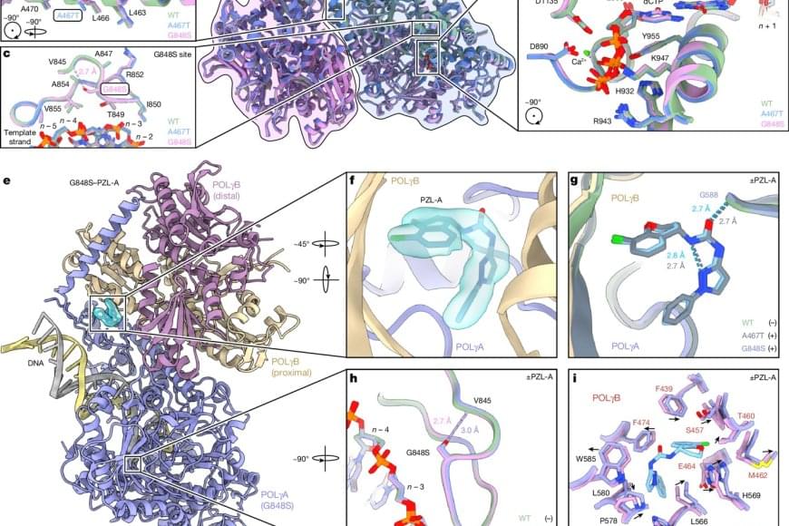
Small molecules restore mutant mitochondrial DNA polymerase activity
Mitochondrial diseases caused by POLG mutations vary in severity. In young children, these diseases can quickly result in brain damage and life-threatening liver problems while others suffer muscle weakness, epilepsy, and organ failure later in childhood. POLG mutations recently received media attention when Prince Frederik of Nassau in Luxembourg died in March 2025 at just 22 years of age.
Gold Nanoparticles Could Restore Vision in People with AMD
Could a tiny dose of gold restore sight? Researchers at Brown University have developed a groundbreaking retinal prosthesis using gold nanoparticles and infrared light to bypass damaged photoreceptors in retinal disorders like macular degeneration.
This minimally invasive method successfully activated the visual system in mice, offering promising early evidence for future clinical applications. Learn how this innovative fusion of nanotechnology and neuroscience could revolutionize treatment for millions suffering from vision loss.
#vision #visionloss #neuroscience #science
Your Brain Can Learn To Ignore Annoying Distractions — Here’s How
The human brain can learn to filter out distracting or disruptive stimuli, such as a bright roadside billboard or a flashing online banner, through repeated exposure. Researchers from Leipzig University and Vrije Universiteit Amsterdam have demonstrated this effect using electroencephalography (EEG), showing that early visual processing in the brain changes with experience. Their findings were recently published in The Journal of Neuroscience.
Distractions tend to become easier to ignore after repeated encounters. This process, known as learned suppression, plays a key role in the visual system and complements our ability to consciously direct attention. In a series of EEG experiments with 24 participants of all genders, the researchers examined how learning affects attention to highly noticeable distractions, particularly when such distractions consistently appear in the same location.