A team from Stanford Medicine has developed AI-powered digital twins of the brain, trained on large datasets of brain activity. These models predict neural responses to new stimuli, offering profound insights into brain function.
People with Alzheimer’s disease may retain their ability to empathize, despite declines in other social abilities, finds a new study led by University College London (UCL) researchers.
The researchers found that people with Alzheimer’s disease scored slightly higher on a measure of empathy than peers of the same age with mild cognitive impairment, despite scoring worse on other measures of social cognition such as recognizing facial emotions and understanding the thoughts of others.
The authors of the study, published in Alzheimer’s & Dementia, say this may be the first time a cognitive domain has been found to improve in dementia.
A brief episode of anxiety may have a bigger influence on a person’s ability to learn what is safe and what is not. Research recently published in npj Science of Learning has used a virtual reality game that involved picking flowers with bees in some of the blossoms that would sting the participant—simulated by a mild electrical stimulation on the hand.
Researchers worked with 70 neurotypical participants between the ages of 20 and 30. Claire Marino, a research assistant in the ZVR Lab, and Pavel Rjabtsenkov, a Neuroscience graduate student at the University of Rochester School of Medicine and Dentistry, were co-first authors of the study.
Their team found that the people who learned to distinguish between the safe and dangerous areas—where the bees were and were not—showed better spatial memory and had lower anxiety, while participants who did not learn the different areas had higher anxiety and heightened fear even in safe areas.
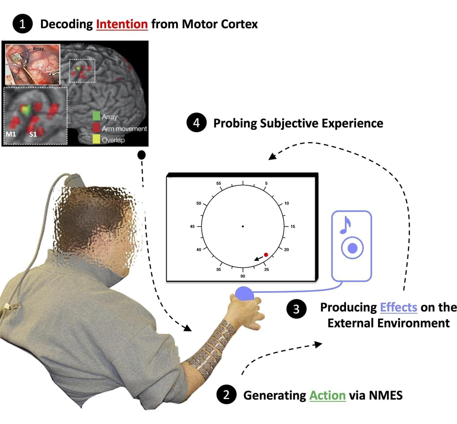
Researchers led by Jean-Paul Noel at the University of Minnesota, United States, have decoupled intentions, actions and their effects by manipulating the brain-machine interface that allows a person with otherwise paralyzed arms and legs to squeeze a ball when they want to.
Published in the open-access journal PLOS Biology, the study reveals temporal binding between intentions and actions, which makes actions seem to happen faster when they are intentional.
Separating intentions from actions was made possible because of a brain-machine interface. The participant was paralyzed with damage to their C4/C5 vertebrae and had 96 electrodes implanted in the hand region of their motor cortex.
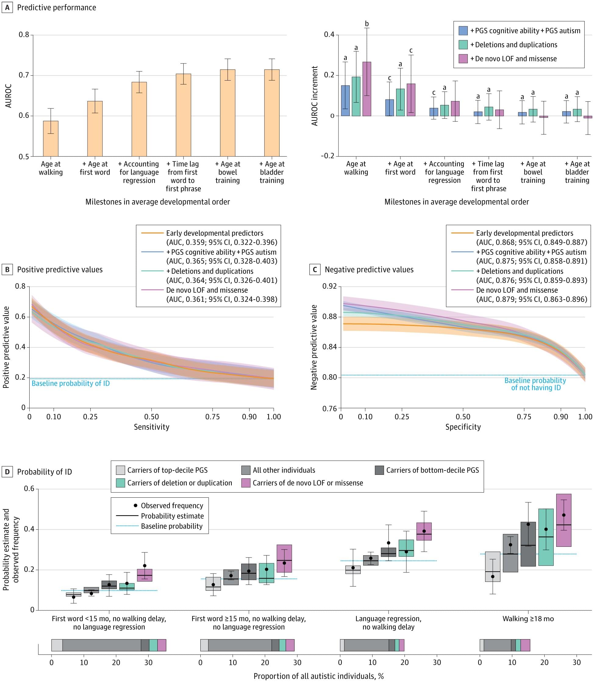
Will a child who’s evaluated for autism later develop an intellectual disability? Can this be accurately predicted? Early-childhood experts in Quebec say they’ve have come up with a better way to find out.
In a study of 5,633 children drawn from three North American cohorts, clinician-researchers affiliated with Université de Montréal developed a new predictive model that combines a wide range of genetic variants with data on each stage of a young child’s development.
Their goal? To obtain reliable information as early as possible to predict the children’s developmental trajectory and thus offer more proactive support to those who may need it—namely, parents trying to better understand and anticipate their child’s needs.
How likely is it that we live in a simulation? Are virtual worlds real?
In this first episode of the 2nd Series we delve into the fascinating topic of virtual reality simulations and the extraordinary possibility that our universe is itself a simulation. For thousands of years some mystical traditions have maintained that the physical world and our separated ‘selves’ are an illusion, and now, only with the development of our own computer simulations and virtual worlds have scientists and philosophers begun to assess the statistical probabilities that our shared reality could in fact be some kind of representation rather than a physical place.
As we become more open to these possibilities, other difficult questions start to come into focus. How can we create a common language to talk about matter and energy, that bridges the simulated and simulating worlds. Who could have created such a simulation? Could it be an artificial intelligence rather than a biological or conscious being? Do we have ethical obligations to the virtual beings we interact with in our virtual worlds and to what extent are those beings and worlds ‘real’? The list is long and mind bending.
Fortunately, to untangle our thoughts on this, we have one of the best known philosophers of all things mind bending in the world, Dr. David Chalmers; who has just released a book ‘Reality+: virtual worlds and the problems of philosophy’ about this very topic. Dr. Chalmers is an Australian philosopher and cognitive scientist specialising in the areas of philosophy of mind and philosophy of language. He is a Professor of Philosophy and Neuroscience at New York University, as well as co-director of NYU’s Center for Mind, Brain and Consciousness. He’s the founder of the ‘Towards a Science of Consciousness Conference’ at which he coined the term in 1994 The Hard Problem of Consciousness, kicking off a renaissance in consciousness studies, which has been increasing in popularity and research output ever since.
Donate here: https://www.chasingconsciousness.net/episodes.
What we discuss in this episode:
00:00 Short Intro.
06:00 Synesthesia.
08:27 The science of knowing the nature of reality.
11:02 The Simulation Hypothesis explained.
15:25 The statistical probability evaluation.
18:00 Knowing for sure is beyond the reaches of science.
19:00 You’d only have to render the part you’re interacting with.
20:00 Clues from physics.
22:00 John Wheeler — ‘It from bit’
23:32 Eugene Wigner: measurement as a conscious observation.
27:00 Information theory as a useful but risky hold-all language tool.
34:30 Virtual realities are real and virtual interactions are meaningful.
37:00 Ethical approaches to Non-player Characters (NPC’s) and their rights.
38:45 Will advanced AI be conscious?
42:45 Is god a hacker in the universe up? Simulation Theology.
44:30 Simulation theory meets the argument for the existence of God from design.
51:00 The Hard problem of consciousness applies to AI too.
55:00 Testing AI’s consciousness with the Turing test.
59:30 Ethical value applied to immoral actions in virtual worlds.
References:
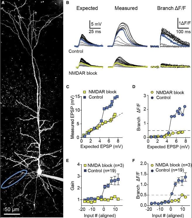
Lafourcade et al. reveal that apical oblique dendrites of retrosplenial cortical L5 neurons exhibit unexpectedly linear integration compared with basal and tuft branches via increased synaptic AMPA: NMDA. Long-range inputs are targeted to these distinct dendritic domains, supporting the idea that single neurons perform a diverse range of subcellular processing.
The pair decided to conduct a clinical trial that could be more compelling. In 12 people with early Alzheimer’s who took 3TC for 6 months, the drug didn’t boost cognitive abilities. But other indicators suggested some benefits, as Frost, Sullivan, and their colleagues revealed last month in npj Dementia. For instance, levels of one key neurodegeneration indicator dipped, suggesting 3TC protects patients’ brain cells. “That was the change I was most excited to see,” Frost says.
Their recent study was the first clinical test of an antitransposon strategy for Alzheimer’s to reach the finish line. But it’s just one of a growing number of trials launched by academic researchers and biotechs to gauge the effects of throttling transposons—so-called jumping genes. These vagrant sequences, some of which are relics of viruses that invaded cells long ago or may even be derived from symbiotic bacteria, make up more than 40% of the human genome but were once seen as largely harmless. However, a variety of evidence from human cell lines, lab animals, and epidemiological studies has implicated their antics in illnesses such as lupus, amyotrophic lateral sclerosis (ALS), Parkinson’s disease, and cancer, as well as in aging.
Encouraging results are trickling in. In 2022, a phase 2 trial determined that 3TC halted tumor growth in some patients with colorectal cancer. Last year, Transposon Therapeutics revealed that a different drug that stymies replication of these sequences slowed one sign of physical decline in people with ALS or another neurodegenerative disease, frontotemporal dementia. “It’s really amazing how quickly the story has developed,” says John Sedivy, a molecular biologist at Brown and the company’s co-founder.