In a unique demonstration of brain implants that incorporate living cells, the devices were able to connect with the brains of live mice.
By Jeremy Hsu
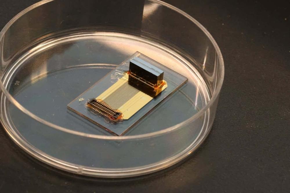
In a unique demonstration of brain implants that incorporate living cells, the devices were able to connect with the brains of live mice.
By Jeremy Hsu

In a recent study published in the journal Nature Aging, researchers identified plasma proteomic biomarkers and dynamic changes associated with brain aging, leveraging a multimodal approach combining brain age gap (BAG) and proteome-wide association analysis.
Background
The global aging population is expected to exceed 1.5 billion individuals aged 65 and above by 2050, highlighting the urgent need to address aging-associated challenges.
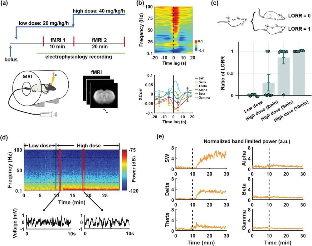
The shift from an awake state to unconsciousness is a phenomenon that has long captured the interest of scientists and philosophers alike, but how it happens has remained a mystery—until now. Through studies on rats, a team of researchers at Penn State has pinpointed the exact moment of loss of consciousness due to anesthesia, mapping what happens in different brain regions during that moment.
The study has implications for humans as well as for other types of loss of consciousness, such as sleep, the researchers said. They published their results in Advanced Science.
“People in the neuroscience field generally understand what happens to a patient who is going under anesthesia at a molecular level,” said corresponding author Nanyin Zhang, the Dorothy Foehr Huck and J. Lloyd Huck Chair in Brain Imaging and professor of biomedical engineering at Penn State.
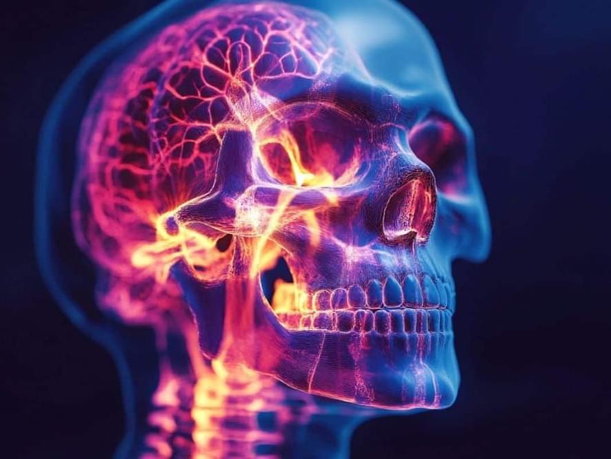
Summary: The dural sinuses and skull bone marrow serve as key communication hubs between the brain’s central immune system and the body’s peripheral immune system. These regions may act as “traffic lights,” allowing immune signals to flow between the brain and body, challenging the traditional view of the blood-brain barrier as an absolute divide.
Researchers found inflammatory activity in these areas correlates with inflammation in both the brain and body, offering new insights into conditions like depression. This discovery could pave the way for innovative treatments targeting these hubs to address immune-related conditions more precisely.
Summary: New research reveals that the cognitive boost from moderate to vigorous exercise lasts up to the next day, enhancing memory performance in adults aged 50 to 83. The study also found that adequate sleep—particularly deep, slow-wave sleep—adds to these benefits.
Conversely, prolonged sedentary time was linked to poorer working memory the following day. These findings highlight the importance of daily physical activity and quality sleep for maintaining cognitive health, especially in older adults.

Synchron has developed a Brain-Computer Interface that uses pre-existing technologies such as the stent and catheter to allow insertion into the brain without the need for open brain surgery.
Read the CNET article for more info:
You Might Not Need Open Brain Surgery to Get Mind Control https://cnet.co/3sZ7k67
0:00 Intro.
0:25 History of Brain Chip Implants.
0:44 About Synchron.
0:54 How Synchron implants the interface.
1:55 How brain patterns transmit signals.
2:50 Risks and Concerns.
3:50 Patients and Clinical Testing.
4:25 Brain Health Monitoring.
5:04 Synchron Switch Price.
Never miss a deal again! See CNET’s browser extension 👉 https://bit.ly/3lO7sOU
Check out CNET’s Amazon Storefront: https://www.amazon.com/shop/cnet?tag=lifeboatfound-20.
Follow us on TikTok: / cnetdotcom.
Follow us on Instagram: / cnet.
Follow us on Twitter: / cnet.
Like us on Facebook: / cnet.
#WhatTheFuture #Synchron #BCI

A new study is shedding light on how stimulating the right bits of the brain can produce dramatic—and seemingly permanent—improvements in the ability of paralysed patients to walk again https://econ.st/3DfZk5L
Photo: NeuroRestore / EPFL 2024
Implanted electrodes allowed one man to climb stairs unaided.
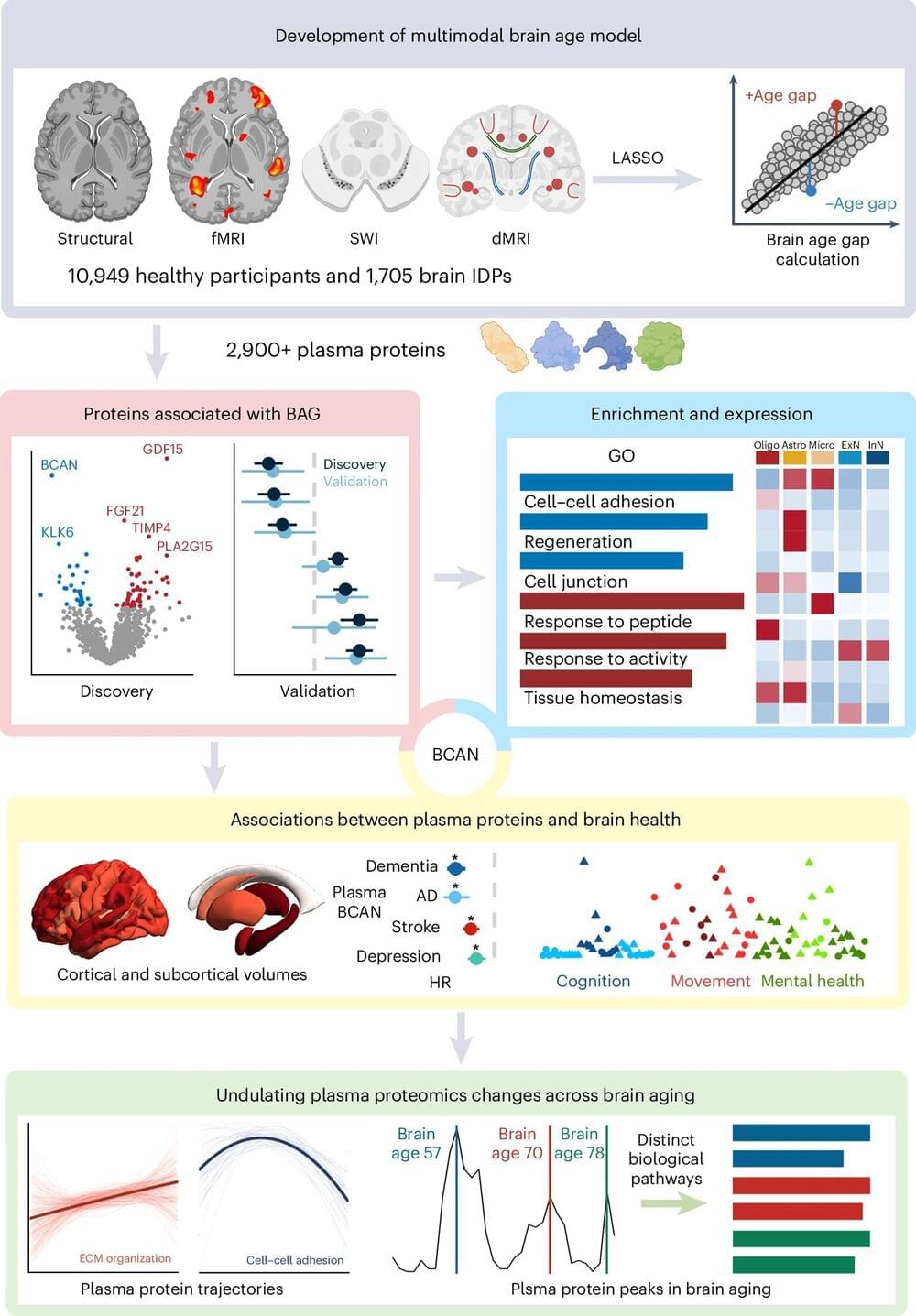
Thirteen proteins linked to brain aging in humans are identified in a Nature Aging paper. Changes in the concentrations of these blood proteins may peak at 57, 70, and 78 years old in humans, and suggest that these ages may be important for potential interventions in the brain aging process.
It is estimated that by 2050 the number of individuals aged 65 years and over will exceed 1.5 billion globally, highlighting the need for a deeper understanding of the aging process—particularly in relation to the brain.
The prevalence of neurodegenerative disorders, such as dementia, is known to increase with aging; however, effective therapies are still limited. The early identification of and intervention in brain aging could help us to prevent such disorders.