New research shows that even mild, prolonged exposure to low oxygen levels can significantly disrupt the blood-brain barrier (BBB) in aging brains.
It’s been thought that T cells would only move into the brain in the case of a serious problem. New work shows otherwise… | Cell And Molecular Biology
Research led by Aarhus University in Denmark reports that individuals with substance use disorders experience a heightened urge to move in response to music with complex rhythms and harmonies.
Long-term use of cocaine and heroin disrupts dopamine signaling in the brain, depleting receptors and diminishing the effects of non-drug stimuli, such as music, to trigger pleasure.
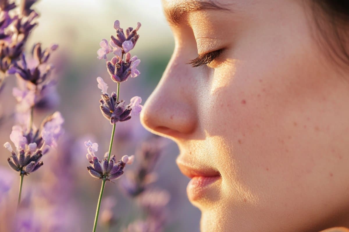
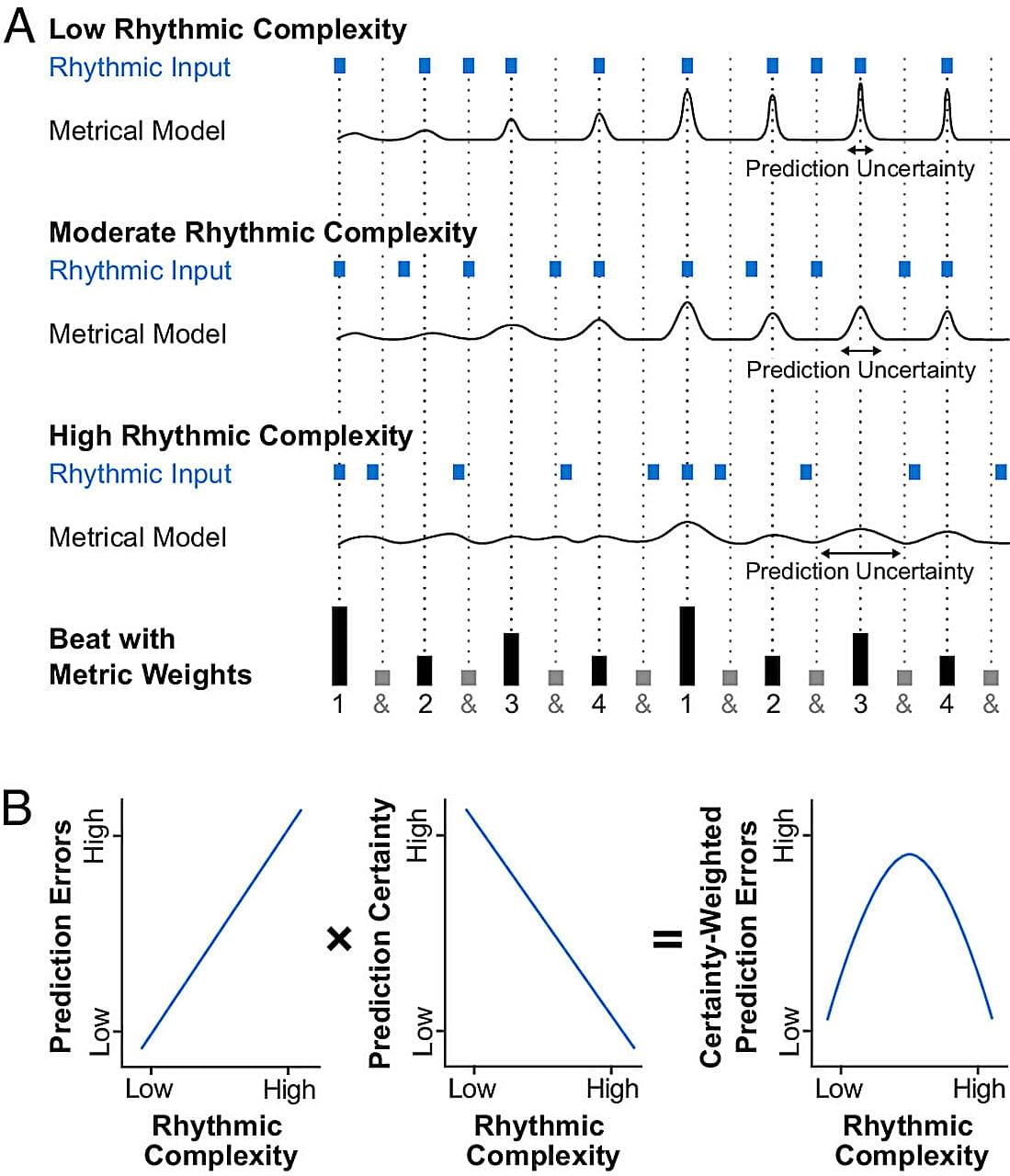
Prior research has shown that music can activate dopaminergic pathways involved in reward, anticipation, and movement. Groove, the pleasurable urge to move to music, follows an inverted-U pattern in healthy listeners, peaking when rhythms fall into a sweet spot of moderate rhythmic complexity. Most people feel the strongest compulsion to move their bodies to the beat when those beats are neither too simple nor too unpredictable.


Humans are social creatures; we live in family groups, socialise with friends, and work with colleagues. Evolutionary psychologist Robin Dunbar’s ‘social brain hypothesis’ suggests that brain size is directly related to social group size in mammals. The bigger the group, the bigger the brain. In this interview with Research Outreach, we find out how Dunbar developed his theory as well as his now famous ‘Dunbar’s number’
Robin Dunbar discusses his eponymous ‘Dunbar’s Number’, primates to people, and why size matters with social groups and evolution.


More and more people are investing their time and energy into longevity — it’s not just living longer, but living happier, healthier and staying productive well past what has been considered “old age.” McKinsey reports that up to 60 percent of consumers across health and wellness markets say that healthy aging is a “top” or “very important” priority. The movement has created a boost in the health and wellness businesses, and to get an overview of the longevity supplements space, we spoke with Dr. Luke Winegard, the Chief Operating Officer at Longevity Method.
Entrepreneur: What is driving the current boom in the longevity supplement market? Dr. Luke Winegard: Growing consumer demand for health and wellness products is creating explosive growth in the longevity supplement market. Scientific advancements and increasing health consciousness are driving this trend, with consumers now focused on “healthspan” — not just how long they live, but how well they live. The pursuit of longevity has moved from being a niche interest of visionaries to becoming mainstream in 2025.
What does “healthspan” mean and why is it important? Healthspan refers to the period of life spent in good health, free from chronic diseases and disabilities. Today’s consumers are concerned not just about adding years to their lives, but making those years healthy, productive, and vital. This represents a cultural shift toward proactive self-optimization where maintaining energy, cognitive sharpness, and resilience is just as important as achieving physical goals.
The brainwaves of people with neuropathic pain show a distinct pattern: more slow theta waves, fewer alpha waves, and more fast, high beta waves, co-lead author Sylvia Gustin, a clinical psychologist and UNSW professor, said in the statement. Her research has investigated changes in the thalamus—a central brain structure that relays sensory and motor signals to the cerebral cortex—associated with nerve pain.
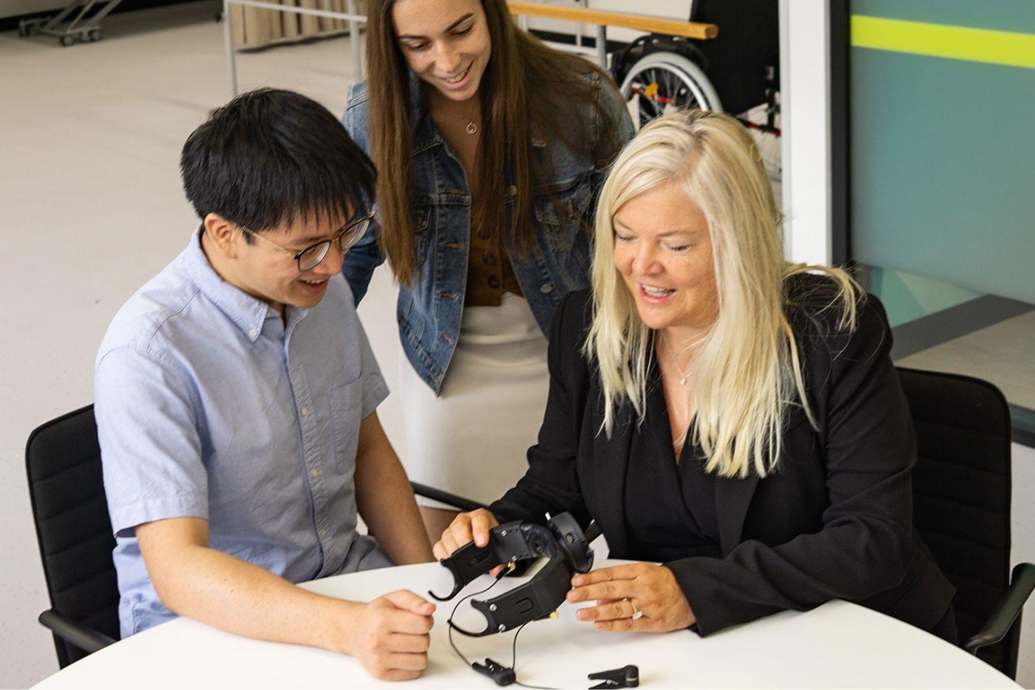
The PainWaive system consists of an electroencephalogram (EEG) headset that records brain activity paired with an app that instructs patients on how to control their brainwaves through neurofeedback games, according to a UNSW statement. Four participants who suffer from corneal neuropathic pain—a condition that causes painful hypersensitivity of the eyes, face, or head—underwent 20 PainWaive sessions over the course of four weeks.
This study offers new hope for drug-free pain treatments, but further trials will need to verify its results.