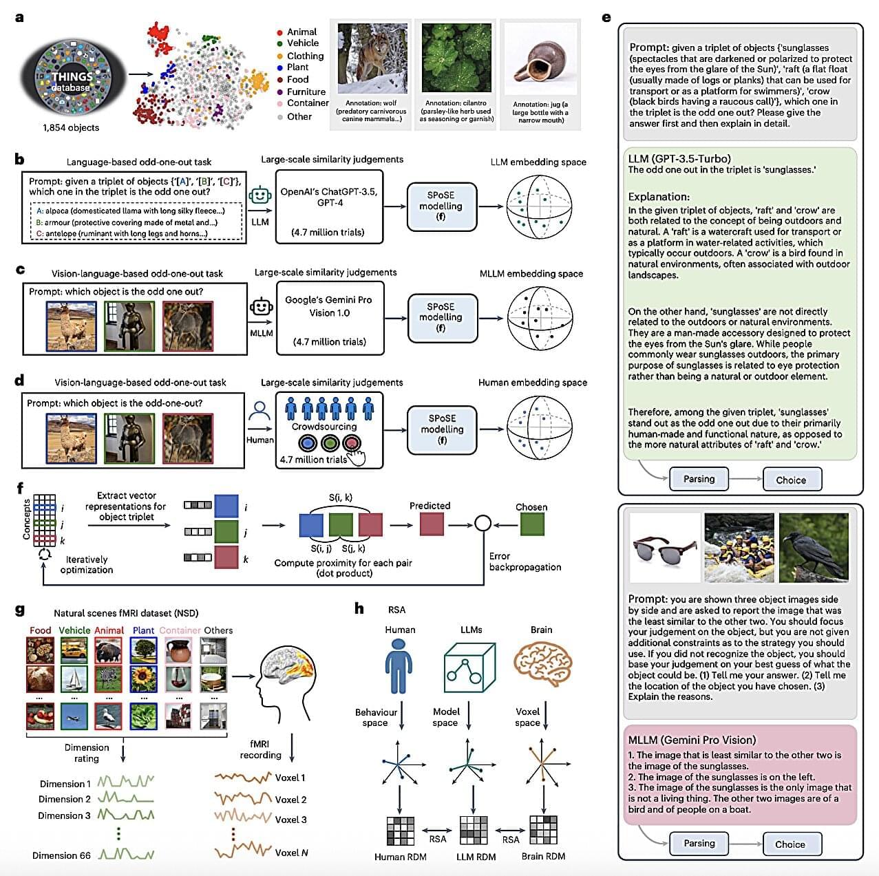
Toggle light / dark theme

We don’t treat Isla’s vision loss as a sad circumstance or as something that is broken in her. It’s so important for us that she knows her vision impairment is not something that makes her less than. If anything, it makes her a stronger, more amazing person, and we couldn’t be prouder of who she is.

Vision impairment is the only symptom she displays of this disease, and we are fighting with everything we have to ensure it stays this way. We were told on diagnosis day that that day was the healthiest Isla would ever be, and that she was at her peak; two years later, and she has continued to defy that, Stockdale added.

The family believes that Isla’s incredible development is a result of the medication, called Miglustat, which she has been taking since November 2022.

Testing time: To see whether the modified VR headset could accurately record brain activity, the researchers had four participants wear it while opening or closing their eyes on command. There is typically an increase in alpha waves in a person’s brain when their eyes are closed, and the researchers could see this increase in the EEG data recorded by their headset.

They also had the participants wear the headset while playing a custom game designed to trigger a type of brain activity that occurs when a person is preparing for an event. As hoped, the researchers were able to see this activity in the recorded EEG data, too.

Looking ahead: The UT Austin team has started filing the paperwork to patent their tech and say they’d be open to working with VR companies to incorporate it into their systems. They don’t say what it costs to make the soft electrodes, so it’s not clear exactly how much consumers or professionals should expect to pay for a modified VR headset.

PHILADELPHIA—Trying to finish your homework while the big game is on TV? “Visual-movement” neurons in the front of your brain can help you stay focused, according to a new study from neuroscientists in the Perelman School of Medicine at the University of Pennsylvania.

In the study, published recently in Neuron, the scientists sought to illuminate the neural mechanism that helps the brain decide whether to focus visual attention on a rewarding task or an alluring distraction. By analyzing neuron activity in animal models as they faced this kind of attentional conflict, the researchers discovered that a pattern of coordinated activity called “beta bursts” in a set of neurons in the lateral prefrontal cortex (LPFC)—a section in the front of the brain responsible for motivation and rewards—appears to have a major role in keeping attention task-focused, essentially by suppressing the influence of the distracting stimulus.

“Our research suggests that while all brains have the ability to focus on a rewarding task and filter out distractions, some are better at it than others,” said senior author Bijan Pesaran PhD, the Robert A Groff II Professor of Neurosurgery at Penn Medicine. “By understanding how our brains process rewarding stimuli, we hope to be able to also understand failures to do so in a variety of cognitive and psychiatric disorders, including attention deficit disorder, schizophrenia, and obsessive-compulsive disorder.”

Every day we encounter circumstances we consider wrong: a starving child, a corrupt politician, an unfaithful partner, a fraudulent scientist. These examples highlight several moral issues, including matters of care, fairness and betrayal. But does anything unite them all?

Philosophers, psychologists and neuroscientists have passionately argued whether moral judgments share something distinctive that separates them from non-moral matters. Moral monists claim that morality is unified by a common characteristic and that all moral issues involve concerns about harm. Pluralists, in contrast, argue that moral judgments are more diverse in nature.

Fascinated by this centuries-old debate, a team of researchers set out to probe the nature of morality using one of moral psychology’s most prolific theories. The group, led by UC Santa Barbara’s René Weber, intensively studied 64 individuals via surveys, interviews and brain imaging on the wrongness of various behaviors.

Similarly, allowing the MyoLegs to flail around for a while in a seemingly aimless fashion gave them better performance with locomotion tasks, as the researchers described in another paper presented at the recent Robotics Science and Systems meeting. Vittorio Caggiano, a Meta researcher on the project who has a background in both AI and neuroscience, says that scientists in the fields of neuroscience and biomechanics are learning from the MyoSuite work. “This fundamental knowledge [of how motor control works] is very generalizable to other systems,” he says. “Once they understand the fundamental mechanics, then they can apply those principles to other areas.”

This year, MyoChallenge 2023 (which will also culminate at the NeurIPS meeting in December) requires teams to use the MyoArm to pick up, manipulate, and accurately place common household objects and to use the MyoLegs to either pursue or evade an opponent in a game of tag.

Emo Todorov, an associate professor of computer science and engineering at the University of Washington, has worked on similar biomechanical models as part of the popular Mujoco physics simulator. (Todorov was not involved with the current Meta research but did oversee Kumar’s doctoral work some years back.) He says that MyoSuite’s focus on learning general representations means that control strategies can be useful for “a whole family of tasks.” He notes that their generalized control strategies are analogous to the neuroscience principle of muscle synergies, in which the nervous system activates groups of muscles at once to build up to larger gestures, thus reducing the computational burden of movement. “MyoSuite is able to construct such representations from first principles,” Todorov says.

Cells hidden in the skull may point to a way to detect, diagnose and treat inflamed brains.

A detailed look at the skull reveals that bone marrow cells there change and are recruited to the brain after injury, possibly traveling through tiny channels connecting the skull and the outer protective layer of the brain. Paired with the discovery that inflammation in the skull is disease-specific, these new findings collectively suggest the skull’s marrow could serve as a target to track and potentially treat neurological disorders involving brain inflammation, researchers report August 9 in Cell.


New observations of skull cell signals and skull tunnels suggest bone marrow there could be used to monitor neurological diseases.

How brain enhancements can correct disabilities is an important question. But not a Closer To Truth question. What happens to personal identity? Would “I” still be me? What about moral standing? Would it change? Could my mind be uploaded? Would “I” live on?

Featuring interviews with Michio Kaku, Antonio Damasio, Leonard Mlodinow, V.S. Ramachandran and David Chalmers.

▶Register for free for subscriber-only exclusives: http://bit.ly/2GXmFsP

Closer To Truth host Robert Lawrence Kuhn takes viewers on an intriguing global journey into cutting-edge labs, magnificent libraries, hidden gardens, and revered sanctuaries in order to discover state-of-the-art ideas and make them real and relevant.

▶Free access to Closer to Truth’s library of 5,000 videos: http://bit.ly/376lkKN

Closer to Truth presents the world’s greatest thinkers exploring humanity’s deepest questions. Discover fundamental issues of existence. Engage new and diverse ways of thinking. Appreciate intense debates. Share your own opinions. Seek your own answers.

Another excellent paper from Johann G. Danzl’s research group. They develop methods that combine novel negative staining techniques, deep learning, and super-resolution STED microscopy or expansion microscopy to facilitate nanoscale-resolution imaging of brain tissue volumes. They also show semi-automated (and some fully automated) segmentation of neuron morphology and identification of synapses. Very cool work and I’m excited to see how it influences connectomics in the future! #brain #neuroscience #imaging #microscopy #neurotech


Mapping fixed brain samples with extracellular labeling and optical microscopy reveals synaptic connections.

The development of your baby’s brain could be influenced by the microbes in their belly, scientists have found.

The trillions of microbes that live inside our guts play essential roles in a range of bodily processes, from digestion to mental health. The gut contains more than 100 million nerve cells—the highest concentration in any part of the body other than the brain. Now, we are also beginning to learn about the roles of these microbes in the earliest stages of our lives.

“The microbiome plays an important role in the early development of several systems, such as the nervous and immune system, as well as providing another layer of protection against pathogens [disease],” Sebastian Hunter, a researcher from the University of British Columbia who led a study on the subject published Wednesday in the scientific journal PLOS One, told Newsweek.