Treating the neurodegenerative disease in its earlier stages is key to slowing cognitive decline. A new study offers hope for the future.

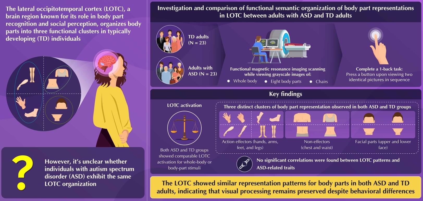
Autism spectrum disorder (ASD), a neurodevelopmental condition, is often associated with difficulties in social communication and repetitive behavior. Previous research reveals that people with ASD often find it challenging to interpret intentions from body language and have difficulty recognizing faces and emotions, which may contribute to their social communication problems.
Scientists speculate that these challenges might arise from differences in how the brain perceives faces and body parts, i.e., how individuals with ASD represent the human body. However, until now, no study had clearly mapped how body parts are represented in the brains of adults with autism or whether that organization differs from normal or typically developing (TD) adults.
In a detailed neuroimaging study involving adults in Japan, researchers addressed this knowledge gap by examining how ASD represents body parts in the brain. This study was published in the journal Imaging Neuroscience. A team of researchers used functional magnetic resonance imaging (fMRI) to compare brain activity patterns in adults with ASD and TD adults as they viewed images of body parts.
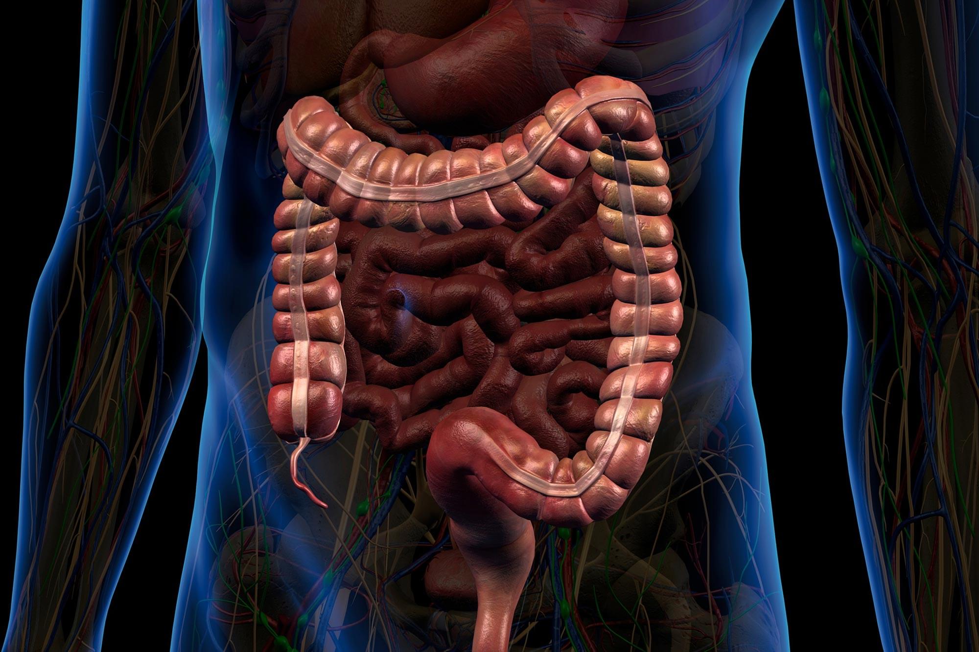
Researchers are gaining a deeper appreciation for the critical role the gastrointestinal (GI) tract plays in maintaining overall health. Beyond its primary responsibilities in digestion, the GI system contributes to the production of hormones, immune cells, and neurotransmitters that influence brain function and emotional well-being.
Because of this, the GI tract contains a wide array of biomarkers that are valuable for diagnosing, tracking, and managing disease—from short-chain fatty acids associated with metabolic syndrome to cytokines linked to inflammation.
However, current technologies fall short when it comes to capturing this biochemical information directly from the GI tract. Existing methods, such as fecal sampling and tissue biopsies, are often invasive, costly, and unable to deliver continuous or comprehensive real-time data throughout the length of the digestive system.
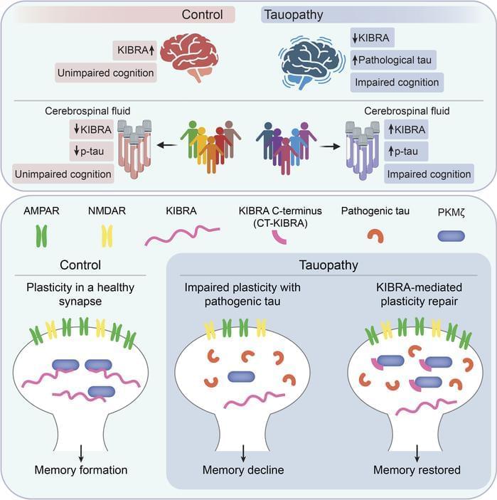
Representing a key mechanism that underlies memory loss in Alzheimer’s disease (AD) and related tauopathies. Here, we found that reduced levels of the memory-associated protein KIdney/BRAin (KIBRA) in the brain and increased KIBRA protein levels in cerebrospinal fluid are associated with cognitive impairment and pathological tau levels in disease. We next defined a mechanism for plasticity repair in vulnerable neurons using the C-terminus of the KIBRA protein (CT-KIBRA). We showed that CT-KIBRA restored plasticity and memory in transgenic mice expressing pathogenic human tau; however, CT-KIBRA did not alter tau levels or prevent tau-induced synapse loss. Instead, we found that CT-KIBRA stabilized the protein kinase Mζ (PKMζ) to maintain synaptic plasticity and memory despite tau-mediated pathogenesis. Thus, our results distinguished KIBRA both as a biomarker of synapse dysfunction and as the foundation for a synapse repair mechanism to reverse cognitive impairment in tauopathy.
1Buck Institute for Research on Aging, Novato, California, USA.
2Memory and Aging Center, Department of Neurology, University of California San Francisco, San Francisco, California, USA.
3Gladstone Institutes, San Francisco, Califoria, USA.
4Weill Institute for Neurosciences, Department of Pathology, University of California San Francisco, San Francisco, California, USA.
In recent years, some scientists and advocates have warned that playing contact sports like football and hockey may increase the risk of brain diseases like Alzheimer’s disease or chronic traumatic encephalopathy (CTE) due to a buildup of a specific protein in the brain.
But a new Northwestern Medicine study of 174 donated brains, including some from former high school and college football players, pumps the brakes on that theory.
“The long and short of it is no, this protein in this specific brain region is not increased in people who played football at the amateur level. It throws a little bit of cold water on the current CTE narrative,” said corresponding author Dr. Rudolph Castellani, professor of pathology at Northwestern University Feinberg School of Medicine and a Northwestern Medicine neuropathologist.

Imagine having a conversation where every gesture and glance feels like a test. You’re juggling eye contact, facial expressions, and tone of voice, all while trying to keep up with the words. You might miss something, or someone might misread you.
In a new study, published in PLOS One, autistic adults describe the intense mental effort it takes to navigate nonverbal communication (NVC).
Researchers reviewed 362 firsthand accounts on the online forum WrongPlanet.net, where autistic adults openly talk about communication challenges. They focused on posts about nonverbal communication—like eye contact, tone of voice, gestures, and facial expressions—and reviewed 26 discussion threads to better understand from autistic adults what it is like to communicate in daily life.

Scientists have discovered how a key protein helps maintain strong connections between brain cells that are crucial for learning and memory.
Results of the study, published in the journal Science Advances, could point the way to new treatments for traumatic brain injuries and diseases, such as Parkinson’s and Alzheimer’s, the scientists said.
Their research, led by a Rutgers University-New Brunswick professor, uncovered a previously unknown role for cypin, a brain protein. Members of the research team found that cypin promotes the presence of tags on specific proteins at synapses, namely the tiny gaps where the brain cells, known as neurons, communicate. The marking helps ensure that the right proteins are in the right place, allowing the synapses to work properly.

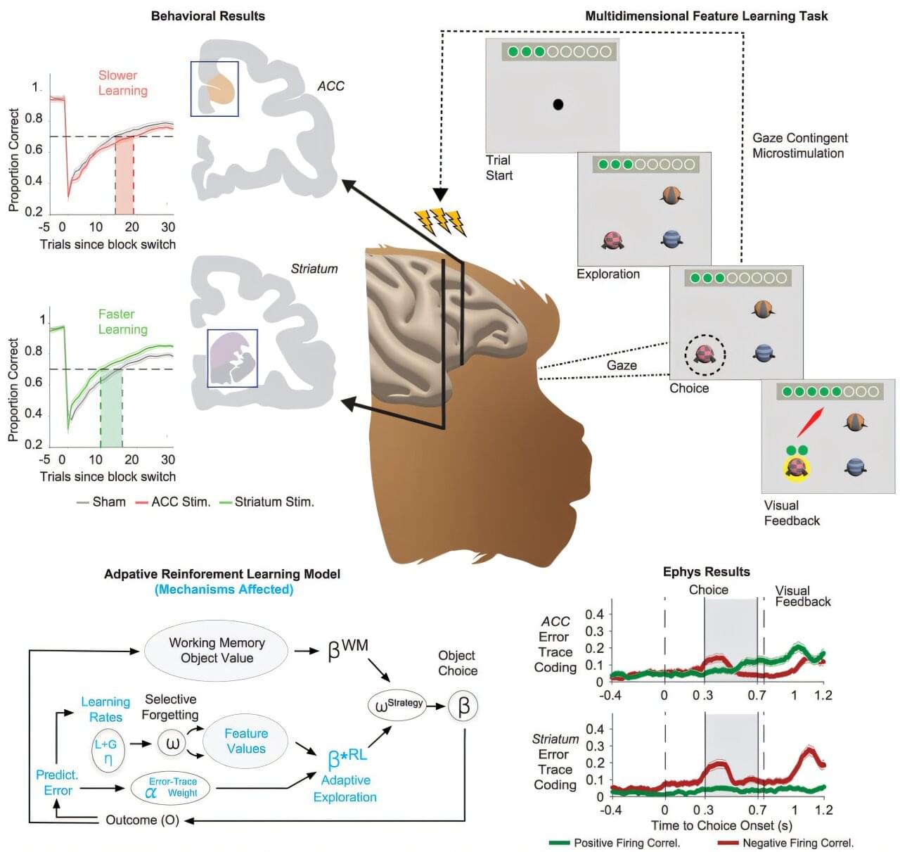
Research led by Thilo Womelsdorf, professor of psychology and biomedical engineering at the Vanderbilt Brain Institute, could revolutionize how brain-computer interfaces are used to treat disorders of memory and cognition.
The study, “Adaptive reinforcement learning is causally supported by anterior cingulate cortex and striatum,” was published June 10, 2025, in the journal Neuron.
According to researchers, neurologists use electrical brain-computer interfaces (BCIs) to help patients with Parkinson’s disease and spinal cord injuries when drugs and other rehabilitative interventions are not efficient. For these disorders, researchers say brain-computer interfaces have become electroceuticals that substitute pharmaceuticals by directly modulating dysfunctional brain signals.