What’s good for your aging gut may also be good for your aging brain. The first study of its kind in twins found that taking daily protein and prebiotic supplements can improve scores on memory tests in people over the age of 60.
Published early last year, the findings are food for thought, especially as the same visual memory and learning test is used to detect early signs of Alzheimer’s disease.
The double-blinded trial involved two cheap plant fiber prebiotics that are available over the counter in numerous nations around the world.
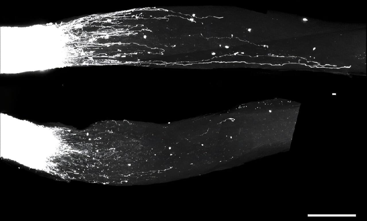
Unlike the brain and spinal cord, peripheral nerve cells, whose long extensions reach the skin and internal organs, are capable of regenerating after injury. This is why injuries to the central nervous system are considered irreversible, while damage to peripheral nerves can, in some cases, heal, even if it takes months or years. Despite decades of research, the mechanisms behind peripheral nerve regeneration remain only partially understood.
In a new study published in Cell, researchers from Prof. Michael (Mike) Fainzilber’s lab at the Weizmann Institute of Science discovered that a family of hundreds of RNA molecules with no known physiological function is essential to nerve regeneration.
Remarkably, the study showed that these molecules can stimulate growth not only in the peripheral nervous system of mice but also in their central nervous system. These findings could pave the way for new treatments for a variety of nerve injuries and neurodegenerative diseases.
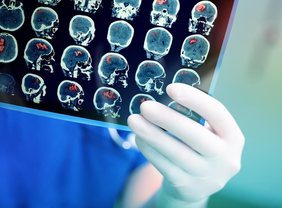
In what experts are calling a “dream come true,” scientists used a recent biochemical discovery to help an 8-year-old boy with a rare genetic condition regain mobility.
Researchers from NYU Langone demonstrated, in a study published in Nature on Wednesday, how a chemical precursor to a commonly available enzyme, CoQ10, can help brain cells overcome a rare genetic condition that severely hobbles cells’ energy production process. Without treatment, the boy’s condition is known to deteriorate rapidly and could be fatal.
NYU Langone researchers have helped an 8-year-old boy regain mobility using an experimental treatment.
Around the world, technology is slowly becoming a part of our bodies. What was once shown only in science fiction movies is now becoming real. For example, in Sweden, thousands of people already have small chips inside their hands. These chips help them open doors, unlock cars, and enter offices—without using keys or cards. These tiny chips make daily life easier and smoother. Now imagine—what if a chip could not only make life easy but also help people with disabilities? This is what Neuralink, a company started by Elon Musk in 2016, is trying to do. Neuralink’s dream is to connect the human brain directly with a computer using a very small chip. Their main aim is to help people who have serious spinal injuries and cannot move. In early trials, Neuralink showed positive results. Some people with paralysis could move a computer cursor or play a chess game—just by thinking. This has given hope to many people who cannot move. But recently, Elon Musk made a new and bold statement that caught the world’s attention. In a post on social media platform X (earlier called Twitter), Musk said that Neuralink’s brain chip could help deaf people hear—even those who were born deaf. He explained that this chip would directly send signals to the part of the brain that understands sound. So, even if a person’s ears do not work, they might still be able to hear. This is different from cochlear implants, which help some deaf people by sending signals to the hearing nerve. Neuralink’s chip would go even deeper—straight to the brain’s hearing area. If successful, this chip could help those who cannot use cochlear implants and give them a new way to experience sound. Elon Musk even said that in the future, such chips might give humans “super-hearing”—allowing them to hear sounds that normal ears cannot hear. However, this is still just an idea. The chip is still being tested. Many technical, safety, and ethical questions are yet to be answered. Also, many Deaf people and experts have said that deafness is not a problem to be “fixed.” For many, deafness is an identity, a language, and a culture. They want to be respected for who they are—not forced to change. At ISH News, we agree with this view. We do not believe that deafness must be “cured.” We also do not support the idea of putting chips inside the body through surgery. But as a news platform made for the Deaf community, we believe it is important to share such news. We want to keep our viewers informed so they can think and talk about these big topics. We are here to provide both sides of the story—the big promises of this new technology, and the serious questions it raises. This way, our community can decide what they think for themselves. The world is now watching to see what Neuralink does next—and whether this brain chip can really change the way people live.
ISH News broadcasts the Daily News and Entertainment online in Deaf-friendly accessible formats which are in Indian Sign Language (ISL), visual images with titles, voice-over and closed-captions. This ensures that we provide equal access to every individual, whilst promoting awareness.
The study, which is set to begin in the third quarter of 2025, will be a randomized, double-blind, placebo-controlled trial involving 40 stroke patients in Europe.
Dr. Nardai, a leading cerebrovascular disease specialist, is currently the Head of the Department of Neurointervention at Semmelweis University in Budapest, Hungary. He previously led a preclinical study published in Experimental Neurology in May 2020, which demonstrated that rats treated with sub-hallucinogenic doses of DMT showed near-complete motor function recovery and smaller infarct volumes compared to untreated control groups. This research provided the basis for Algernon’s clinical investigation into DMT as a potential neuroprotective agent for stroke recovery.
“The primary endpoint of the planned Phase 2a study will be safety,” said Dr. Nardai in the news release. “However, stroke clinicians worldwide will also be watching for positive signals regarding lesion volume, biomarkers, motor function, cognitive function, depression, and mortality.””
Algernon Pharmaceuticals Inc. (AGN: CSE; AGNPF: OTCQB; AGW0:XFRA) subsidiary Algernon NeuroScience has appointed Dr. Sandor Nardai as Principal Investigator for its upcoming Phase 2a DMT stroke study. Find out how this trial could reshape stroke recovery research.
Discover how MitoQ reverses vascular aging by 15–20 years! Chief Scientific Officer Dr. Mitchell reveals research on mitochondrial health, showing 42% improvement in blood flow and superior results vs CoQ10 for longevity. Some links are affiliate links so we will earn a commission when they are used to purchase products.
If you would like to support our channel please consider joining our Patreon / modernhealthspan. MitoQ https://tinyurl.com/5n93fm3f MitoQ Pure https://tinyurl.com/3hxy9s7x. Stemregen 15% discount Code MODERN https://tinyurl.com/45z968yr (Only available in the US) AX3 Life Astaxanthin 20% discount code MODERN20 https://tinyurl.com/2t3w26nw. All Muse devices 15% discount code MODERN https://choosemuse.com/MODERN Renue By Science 10% discount code MHS: https://tinyurl.com/bdew4bfs. NMN Powder https://tinyurl.com/syc7rwkh. DoNotAge 10% discount code MHS https://tinyurl.com/6dbvhv87 D3/K2 https://tinyurl.com/528z26b7 NMN https://tinyurl.com/wyzj2f3d. n1o1 Nitric Oxide 10% discount with code Modern https://tidd.ly/3IczGRW 15% off with code PRIME15 until July 12 n1o1 Nitric Oxide Lozenges https://tidd.ly/4eaJTdw. Age-Defiance Face Cream https://tidd.ly/4eaJTdw. Wellness Extract 10% discount Code MODERNWE Geranylgeraniol Essential http://wellnessextract.com/RICHARDWE Delta Gold Vit E Oxford Healthspan Spermidine 15% Code MHS Original https://tinyurl.com/hrxfnzpn, Gluten Free https://tinyurl.com/2s39pkzv. Nuchido Time+ 20% discount of first purchase with code MODERN20 https://nuchido.com/MODERN Pendulum 20% Discount Code HEALTHSPAN Akkermansia https://pendulumtherapeutics.sjv.io/b… Daily https://pendulumtherapeutics.sjv.io/N… OmegaQuant 5% discount Code MODERN https://omegaquant.com/shop/ OneSkin 15% Discount: Code MODERN OS-01 Face https://oneskin.pxf.io/Z6Yg0K In this comprehensive interview, Dr. Mitchell, Chief Scientific Officer at MitoQ, breaks down the revolutionary science behind mitochondrial-targeted antioxidants and their impact on aging and disease prevention. *Key Topics Covered:* • How MitoQ delivers 90% bioavailability to mitochondria vs 10% for regular CoQ10 • Clinical study showing 42% improvement in flow-mediated dilation (vascular function) • Why mitochondrial dysfunction is the root cause of aging diseases • Exercise performance benefits: improved VO2 max, peak power, and recovery • Brain health and cognitive function improvements • Cardiovascular benefits: reduced oxidized LDL, improved artery flexibility • Immune system support and inflammation reduction • Diabetes prevention through improved glucose metabolism • Proper dosing protocols and supplement combinations *Research Highlights:* ✓ 25+ clinical studies and 800+ research papers ✓ Reverses vascular aging by 15–20 years according to leading researcher Doug Seals ✓ Professional cyclists and elite athletes using MitoQ for performance ✓ Ongoing trials for frailty, cognitive function, and neurovascular health ✓ 300+ independent researchers studying MitoQ applications Learn why targeting mitochondria may be the key to preventing age-related diseases and optimizing healthspan. Discover the science-backed approach to cellular energy and antioxidant protection that’s changing how we think about aging. ⏲️Chapters 0:00 The problem with mitochondria 3:34 How Mitochondria Dysfunction Occurs with Aging 5:43 Natural Protective Mechanisms and CoQ10 7:38 The Invention of MitoQ — Superior Mitochondrial Targeting 11:15 How MitoQ Differs from CoQ10 13:31 MitoQ vs CoQ10 — Research Comparisons 17:52 Cardiovascular Benefits and Flow-Mediated Dilation 23:32 Heart Muscle Function and Immune System Effects 28:16 Exercise Performance and V2 Max Improvements 32:04 Research Funding and Collaborative Programs 36:11 Diabetes Prevention and Glucose Metabolism 39:02 Brain Health and Cognitive Benefits 44:23 Dosing Protocol and Administration Guidelines 48:04 Supplement Combinations and Future Directions 🌐Links in this video MitoQ Home page https://www.mitoq.com/ Mitochondrial Collaborative Research Program https://www.mcrp.dev/ *************************************** Health claims Disclosure: Information provided on this video is not a substitute for direct, individual medical treatment or advice. Please consult with your doctor first. Products or services mentioned in this video are not a recommendation. Audio Copyright Disclaimer Please note that we have full authorization to the music that we used in our videos as they were created using the service WeVideo which provides the rights to the music. The rights are detailed in the terms of use that can be reviewed here https://www.wevideo.com/terms-of-use and any following inquiries should be addressed to [email protected]. ************************************************ #MitoQ #Mitochondria #Longevity. Metabolic Daily https://pendulumtherapeutics.sjv.io/N… OmegaQuant 5% discount Code MODERN https://omegaquant.com/shop/ OneSkin 15% Discount: Code MODERN OS-01 Face https://oneskin.pxf.io/Z6Yg0K
In this comprehensive interview, Dr. Mitchell, Chief Scientific Officer at MitoQ, breaks down the revolutionary science behind mitochondrial-targeted antioxidants and their impact on aging and disease prevention.
*Key Topics Covered:* • How MitoQ delivers 90% bioavailability to mitochondria vs 10% for regular CoQ10 • Clinical study showing 42% improvement in flow-mediated dilation (vascular function) • Why mitochondrial dysfunction is the root cause of aging diseases. • Exercise performance benefits: improved VO2 max, peak power, and recovery. • Brain health and cognitive function improvements. • Cardiovascular benefits: reduced oxidized LDL, improved artery flexibility. • Immune system support and inflammation reduction. • Diabetes prevention through improved glucose metabolism. • Proper dosing protocols and supplement combinations.
*Research Highlights:* ✓ 25+ clinical studies and 800+ research papers. ✓ Reverses vascular aging by 15–20 years according to leading researcher Doug Seals. ✓ Professional cyclists and elite athletes using MitoQ for performance. ✓ Ongoing trials for frailty, cognitive function, and neurovascular health. ✓ 300+ independent researchers studying MitoQ applications.
Learn why targeting mitochondria may be the key to preventing age-related diseases and optimizing healthspan. Discover the science-backed approach to cellular energy and antioxidant protection that’s changing how we think about aging.
Researchers at Princeton University and the Simons Foundation have identified four clinically and biologically distinct subtypes of autism, marking a transformative step in understanding the condition’s genetic underpinnings and potential for personalized care.
Analyzing data from over 5,000 children in SPARK, an autism cohort study, the researchers used a computational model to group individuals based on their combinations of traits.
The team used a “person-centered” approach that considered a broad range of over 230 traits in each individual, from social interactions to repetitive behaviors to developmental milestones, rather than searching for genetic links to single traits.
Clinical studies suggest the therapeutic potential of psychedelics, including ayahuasca, DMT, psilocybin, and LSD, in stress-related disorders. These substan…
If you regularly experience headaches, dizziness, balance problems and blurred vision, our Neanderthal cousins could be to blame.
These are common symptoms of Chiari malformations, structural defects in which the lower part of the brain extends into the spinal cord. People with this condition have skulls shaped like those of our ancient relatives, leading to a hypothesis (known as the Archaic Homo Introgression Hypothesis) that it may be a genetic legacy from interbreeding between modern humans and Neanderthals.
To investigate this, Kimberly Plomp of the University of the Philippines Diliman and colleagues zeroed in on Chiari 1, the mildest form of the condition, which affects around 1 in 100 people.
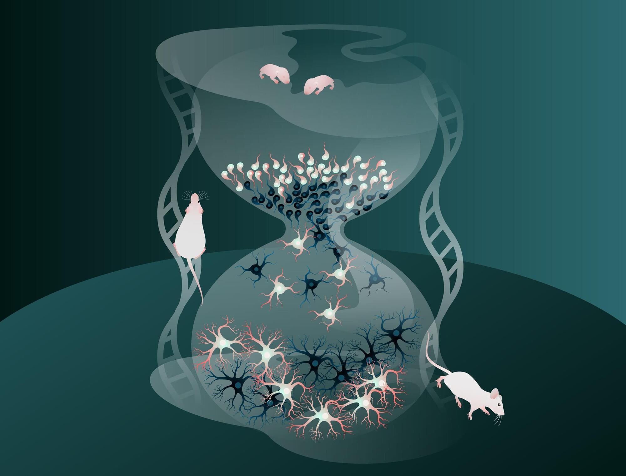
The human brain is made up of billions of nerve cells (neurons) that communicate with each other in vast, interconnected networks. For the brain to function reliably, there must be a fine balance between two types of signals: Excitatory neurons that pass on information and increase activity, and inhibitory neurons that limit activity and prevent other neurons from becoming too active or firing out of control. This balance between excitation and inhibition is essential for a healthy, stable brain.
Inhibitory neurons are generated during brain development through the division of progenitor cells—immature cells not yet specialized but already on the path to becoming neurons. A new study, led by researchers at the Max Planck Institute for Biological Intelligence, has uncovered a surprising feature of brain development based on findings in mice: During this essential process, cells born later in development mature much more quickly than those produced earlier.
The findings are published in the journal Nature Neuroscience.