Could invisible mold toxins be the hidden reason behind fatigue, brain fog, or stubborn inflammation?

This is a ~1 hour talk by Michael Timothy Bennett (https://michaeltimothybennett.com/) on his ideas around computation and consciousness.
Direct link to the thesis preprint: https://osf.io/preprints/thesiscommons/wehmg_v1?view_only=
YT: https://www.youtube.com/@michaeltimothybennett.
X: https://twitter.com/MiTiBennett
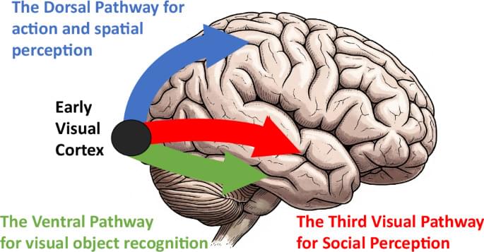
Recent evidence suggests the existence of a neural pathway specialized for social perception projecting between the well-established “what” and “where” pathways. A new study of neuropsychological patients demonstrates that this social pathway is causally essential for recognizing dynamic facial expressions.
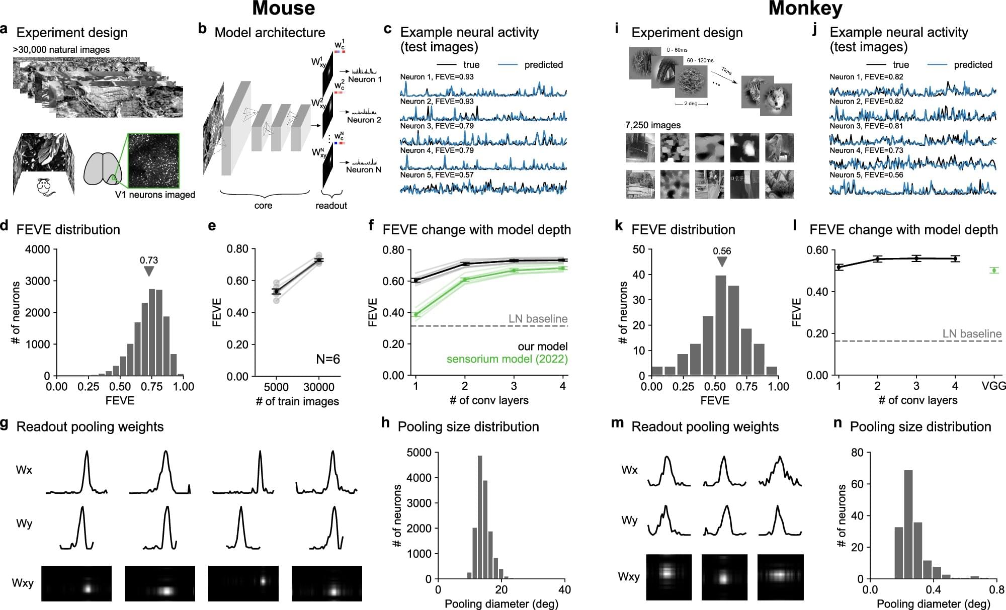
Neuroscientists want to understand how individual neurons encode information that allows us to distinguish objects, like telling a leaf apart from a rock. But they have struggled to build computational models that are simple enough to allow them to understand what individual neurons are doing.
To address this challenge, researchers in the Stringer and Pachitariu labs at Janelia set out to create a simpler model to explain what’s going on in the primary visual cortex —the first stop in the brain for visual data. Their paper is published in the journal Nature Communications.
“We are trying to build a model that can predict the visual responses of each individual neuron,” says Fengtong Du, a graduate student in the Stringer Lab who led the new research.

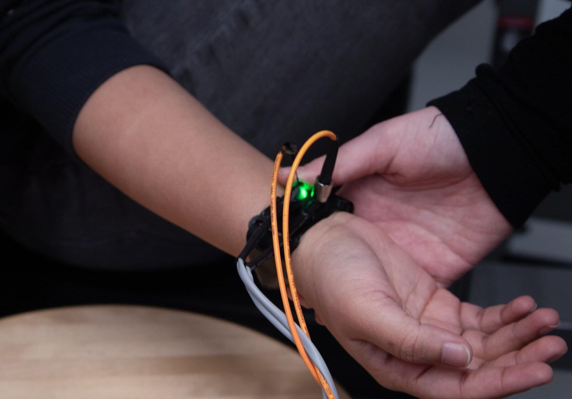
Researchers have shown, for the first time, that speckle contrast optical spectroscopy (SCOS) can be used for cuffless blood pressure monitoring. The new technology could improve early detection and management of hypertension.
“Hypertension affects nearly half of all adults in the US and is the leading cause of cardiovascular disease,” said Ariane Garrett, a doctoral student in Darren Roblyer’s lab at Boston University. “This research is a step toward a wearable device that would let people monitor their blood pressure any time, without a cuff.”
SCOS is a noninvasive imaging technique that measures blood flow by analyzing speckle patterns formed by coherent light scattering from cells and tissue. While it has been used for other applications such as brain and tissue monitoring, this is one of the first studies to explore how SCOS signals relate to blood pressure.
Have you heard about the crazy guys who bought an entire tower to convert it into a vertical village? Yes, that’s us.
Do you want to walk the 16-floor tower and explore the space? Still on the fence, if you should become a citizen? Do you have questions about how you can get involved and co-create? Wanna hear updates on what happened in the last 2 weeks? This event is for you! 👩🚀
About us: We are transforming a 16-floor tower in the heart of San Francisco into a self-governed vertical village —a hub for frontier technologies and creative arts. 8 themed floors will be dedicated to creating tier-one labs, spanning AI, Ethereum, biotech, neuroscience, longevity, robotics, human flourishing, and arts & music. These floors will house innovators and creators pushing the boundaries of human potential in a post-AI-singularity world.
In an exclusive interview, Noland Arbaugh discusses becoming the first person to receive Neuralink’s brain-computer interface, The Link.
I believe that dna will be able to answer just about all our genetic coding questions so much that it will lead to even better breakthroughs in the future and use hardly any energy. I believe also that the master algorithm can eventually be derived from DNA as dna seems already a perfect master algorithm for human beings where human beings are the key to all future progress. I say this as quantum computing is still not stable but we already know that dna computers seem already a masterpiece already especially even organoids of the human brain. Really it becomes really quite simple as even the quantum realm is unstable but dna computers that are quantum would stabilize this currently unstable realm.
Riera Aroche, R., Ortiz García, Y.M., Martínez Arellano, M.A. et al. DNA as a perfect quantum computer based on the quantum physics principles. Sci Rep 14, 11,636 (2024). https://doi.org/10.1038/s41598-024-62539-5