Researchers have discovered the brain uses fat the same way muscles do — as an alternative fuel to glucose — going against nearly a century of accepted science.

Macquarie University new study could hold the key to developing therapies for devastating age-related diseases such as motor neuron disease (MND), Alzheimer’s disease, and Parkinson’s disease.
The research at Macquarie University conducted by neurobiologist, Dr. Sina Shadfar and colleagues in the Motor Neuron Disease Research Centre, reveals a protein called protein disulphide isomerase (PDI) helps repair serious deoxyribonucleic acid (DNA) damage. This breakthrough opens new possibilities for therapies aimed at boosting the body’s ability to fix its own DNA, a process that becomes less efficient as we age.
Dr. Shadfar, Associate, Macquarie Medical School stated “Brain cells are very vulnerable. Unlike skin or blood cells, they don’t divide or renew so any damage that builds up in them stays and if the damage isn’t repaired, it can eventually lead to the death of these critical cells.”

Bipolar disorder is a mental health condition characterized by extreme mood swings, with alternating periods of depression and manic episodes. Past research suggests that bipolar disorder has a strong genetic component and is among the most heritable psychiatric disorders.
To better understand the genetic factors that increase the risk of developing this mental health disorder, neuroscientists and geneticists have carried out various genome-wide association studies (GWAS). These are essentially studies aimed at identifying specific regions of the human genome that are linked with an increased risk of having bipolar disorder, also referred to as bipolar risk loci.
While earlier works have identified many of these regions, causal single nucleotide polymorphisms (SNPs) for the disorder are largely unknown. These are essentially genetic variants that primarily contribute to bipolar disorder risk, as opposed to just being mere markers of it.

Acute brain injuries (ABIs) pose a substantial global burden, demanding effective prognostic indicators for outcomes. This study explores the potential of urinary p75 neurotrophin receptor (p75NTR) concentration as a prognostic biomarker, particularly in relation to unfavorable outcomes. The study involved 46 ABI patients, comprising sub-cohorts of aneurysmal subarachnoid hemorrhage, ischemic stroke, and traumatic brain injury. Furthermore, we had four healthy controls. Samples were systematically collected from patients treated at the University Hospital of Turku between 2017 and 2019, at early (1.50 ± 0.70 days) and late (9.17 ± 3.40 days) post-admission time points. Urinary p75NTR levels, measured by ELISA and normalized to creatinine, were compared against patients’ outcomes using the modified Rankin Scale (mRS). Early urine samples showed no significant p75NTR concentration difference between favorable and unfavorable mRS groups. In contrast, late samples exhibited a statistically significant increase in p75NTR concentrations in the unfavorable group (p = 0.033), demonstrating good prognostic accuracy (AUC = 70.9%, 95% CI = 53–89%, p = 0.03). Assessment of p75NTR concentration changes over time revealed no significant variation in the favorable group (p = 0.992) but a significant increase in the unfavorable group (p = 0.009). Moreover, p75NTR concentration was significantly higher in ABI patients (mean ± SD 40.49 ± 28.83–65.85 ± 35.04 ng/mg) compared to healthy controls (mean ± SD 0.54 ± 0.44 ng/mg), irrespective of sampling time or outcome (p < 0.0001). In conclusion, late urinary p75NTR concentrations emerged as a potential prognostic biomarker for ABIs, showing increased levels associated with unfavorable outcomes regardless of the specific type of brain injury. While early samples exhibited no significant differences, the observed late increases emphasize the time-dependent nature of this potential biomarker. Further validation in larger patient cohorts is crucial, highlighting the need for additional research to establish p75NTR as a reliable prognostic biomarker across various ABIs. Additionally, its potential role as a diagnostic biomarker warrants exploration.

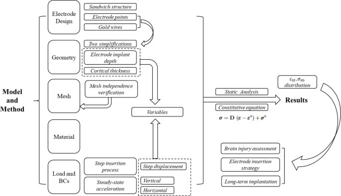
Peng, L., Wang, L., Wu, S. et al. Sci Rep 15, 19,938 (2025). https://doi.org/10.1038/s41598-025-04737-3
It’s 3:43 AM. Sirens are howling. Your phone lights up: DEFCON 1. Multiple ICBMs inbound.
World War 3 has just begun.
Would you know what to do in the first minutes? Most people freeze. This guide is for those who act.
In this video, we walk you through the real first steps to take if global war breaks out — not theory, not panic, but practical survival strategy for the first 24 hours: from identifying if you’re in a high-risk zone, to securing water and food, to communicating with loved ones when the grid is down.
💥 Whether it’s a nuclear attack, an EMP, or a cyber blitz — this is what you need to know before it’s too late.
Topics we cover include: