From mini-brains and spider-inspired gloves to edible wolf apple coatings and microplastic-filled retinas, scientists are transforming creepy concepts into life-improving innovations. Lab-grown brain organoids could replace animal testing, web-slinging gloves can spin instant wound dressings, and wolf apple starch may keep veggies fresh longer. Meanwhile, the discovery of microplastics in human eyes reveals a haunting truth about our environment’s reach inside us.
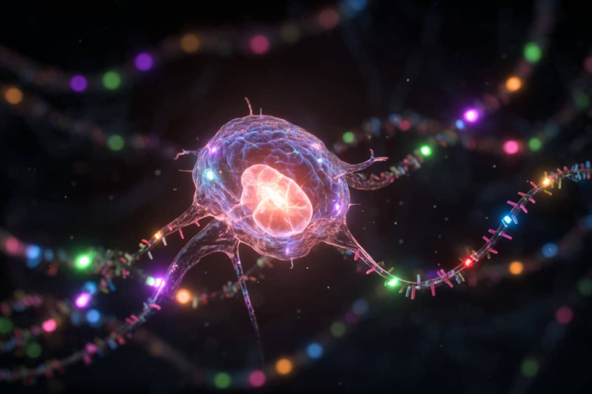
Lab-Grown “Mini-Brains” Offer New Insight into the Human Mind
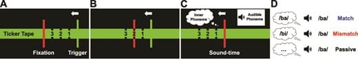
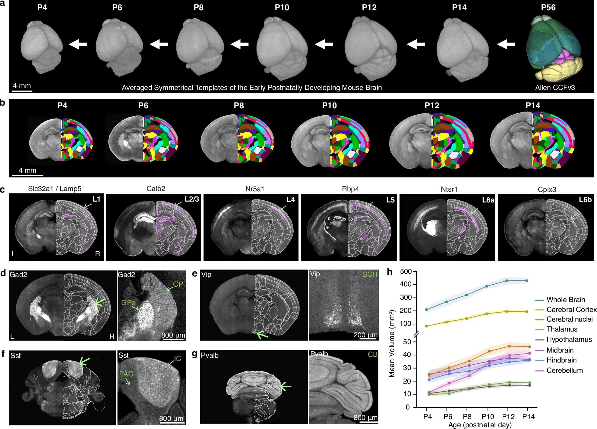
Scientists writing in ACS Sensors have successfully grown a small brain organoid in a petri dish, creating a powerful new tool for studying how nerve cells interact without the use of animal testing. Over two years, human nerve cells multiplied and organized themselves into a three-dimensional “mini-brain” that displayed electrical activity similar to real brain tissue. Researchers say this breakthrough could help scientists better understand how the human brain communicates and functions—or, as they joke, provide “a lab-grown lunch option for zombies.”