Study maps five major eras of brain wiring from birth to old age, revealing the key turning points that shape how we learn, think, and age.
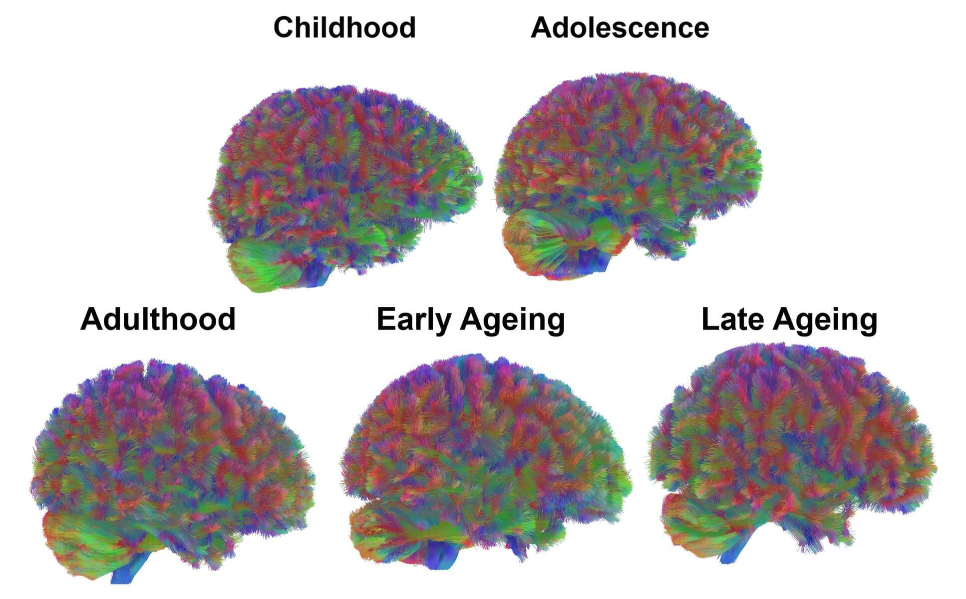
As we get older, our brains start to change in ways that make them increasingly vulnerable to disease – and a detailed new study of these changes points to a way some of this wear and tear might be prevented or reversed.
Researchers from the Leibniz Institute on Aging – Fritz Lipmann Institute in Germany used mass spectrometry to analyze the balance of brain proteins in both young and old mice, finding differences in a process called ubiquitylation as the animals aged.
Ubiquitylation adds chemical tags to proteins, telling the brain which of these busy molecules are past their peak and should be recycled. In older mouse brains, the ubiquitylation tags really start to pile up on certain proteins.
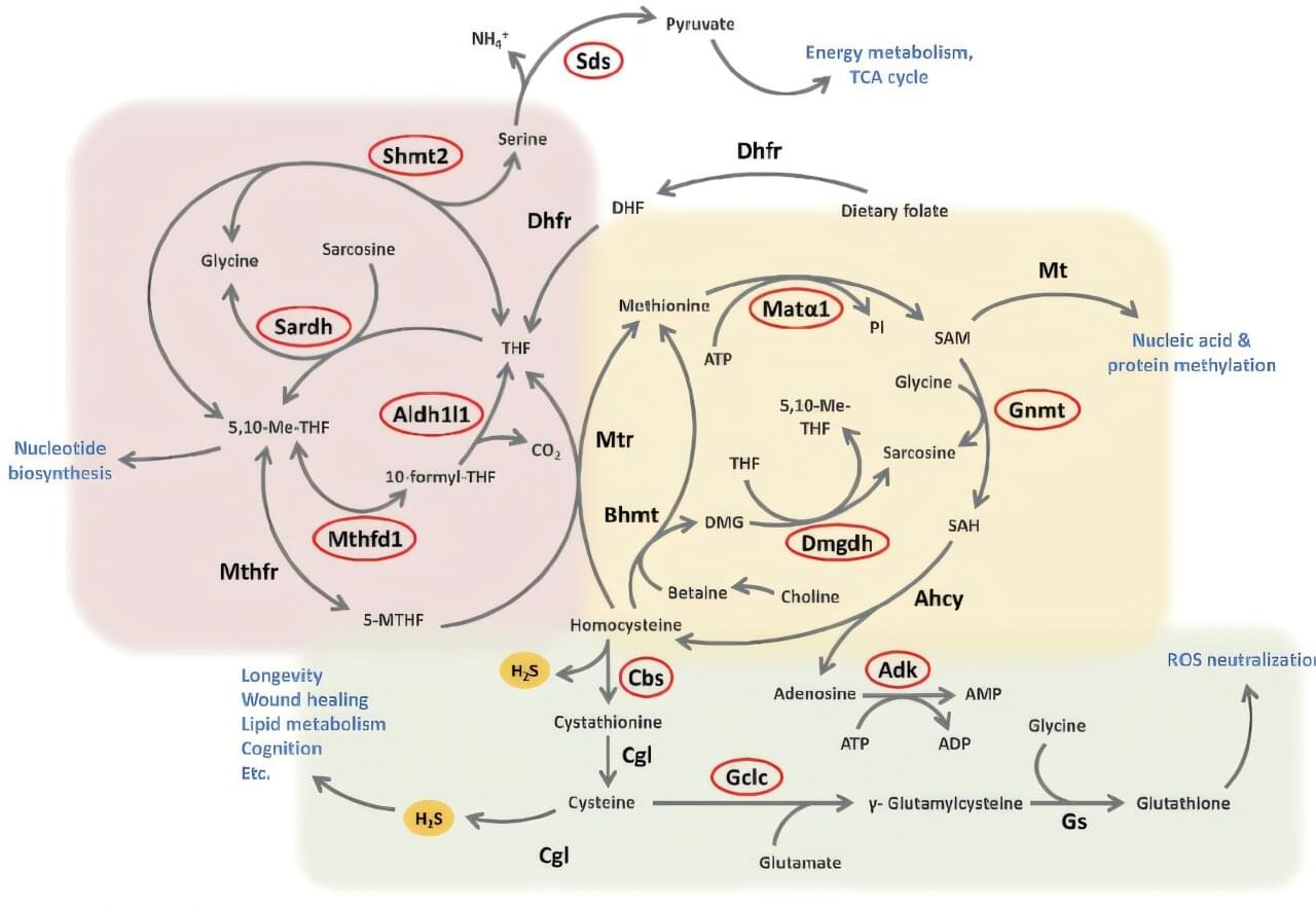
Researchers at Bar-Ilan University have discovered how the longevity-associated protein Sirt6 orchestrates a delicate molecular balancing act that protects the body from age-related decline and disease. The new findings, just published in the Proceedings of the National Academy of Sciences, reveal how Sirt6 preserves health during aging and may pave the way for therapies that promote a longer, healthier life.
Sirt6, often described as a master regulator of aging, is known for its powerful protective effects against age-related diseases such as cancer, diabetes, inflammation, and frailty. Its impact closely resembles that of calorie restriction, a dietary regimen proven in animals to extend lifespan and enhance the body’s natural repair and healing mechanisms.
Calorie restriction—eating fewer calories without malnutrition—has long been known to improve health and extend lifespan. One of its key effects is to increase the body’s production of hydrogen sulfide (H2S), a tiny gas molecule that supports wound healing, heart health, and brain function. This new study found that as we age, H2S levels naturally decline, weakening these protective benefits.
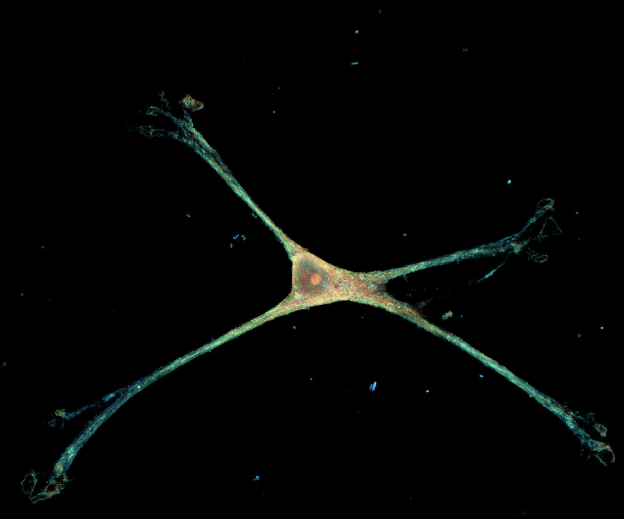
Scientists have engineered a nanowire platform that mimics brain tissue to study astrocytes, the star-shaped cells critical for brain health, for the first time in their natural state.
Astrocytes are the brain’s most abundant and mysterious cells, responsible for regulating communication between neurons and helping to maintain the blood-brain barrier. They are also highly dynamic shape-shifters, something they do not do on typical petri dishes, leaving major gaps in our understanding of how they operate.
“Frustratingly, little is known about the stunning diversity of astrocyte morphology and we also don’t know much about the molecular machinery behind these shape shifts,” said co-senior author Ishan Barman, a Johns Hopkins University bioengineer. “They won’t take on these shapes on glass, so the question for us was how do we replicate the in vivo shape but in vitro?”
Summary: Researchers at Ruhr University Bochum explore why consciousness evolved and why different species developed it in distinct ways. By comparing humans with birds, they show that complex awareness may arise through different neural architectures yet serve similar purposes.
New research examines why consciousness evolved by comparing humans with birds.
What evolutionary purpose does consciousness serve, and what insights can birds offer about its origins? These questions are at the heart of two new studies from researchers at Ruhr University Bochum.
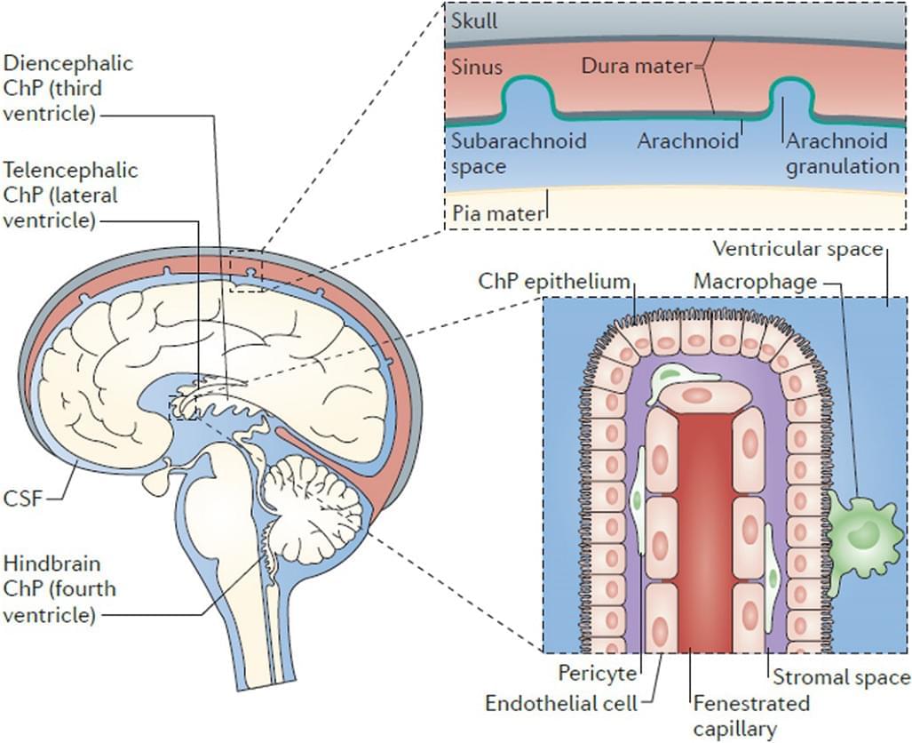
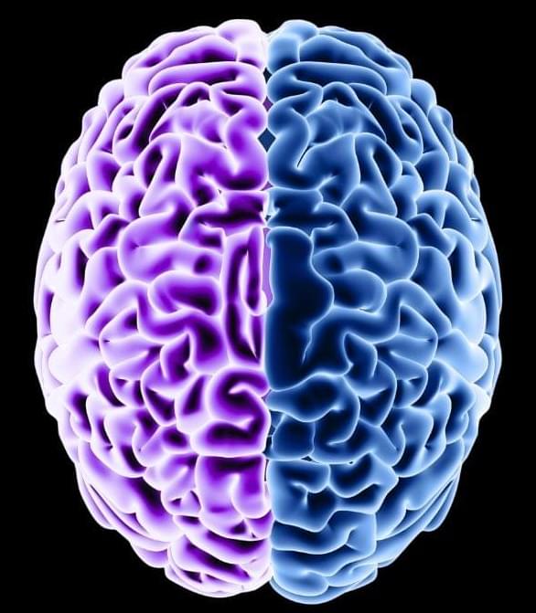
The accompanying diagram presents a comprehensive anatomical overview of the human brain, integrating both lateral surface morphology and a midsagittal section to illustrate the spatial organization of cortical and subcortical structures. Major gyri, sulci, and lobar divisions are delineated alongside deep nuclei, commissural pathways, and the ventricular system. The transparent rendering of the ventricles highlights their relationship to surrounding neural tissue and emphasizes the topology of cerebrospinal fluid pathways. This visualization serves as a structural reference point for understanding functional domains such as sensorimotor processing, higher-order cognition, limbic integration, and autonomic regulation. Collectively, the diagram provides a detailed framework for interpreting neuroanatomical connectivity and its relevance to neural function.
#study:
Cerebrospinal Fluid Mechanics and Its Coupling to Cerebrovascular Dynamics: https://www.annualreviews.org/content/journals/10.1146/annur…#45;034321
CSF dynamics throughout the ventricular system using 4D flow MRI: associations to arterial pulsatility, ventricular volumes, and age: https://link.springer.com/article/10.1186/s12987-024-00570-4
Fundamental functional differences between gyri and sulci: implications for brain function, cognition, and behavior: https://pubmed.ncbi.nlm.nih.gov/38665307/?utm_source=chatgpt.com.
Brain ventricles as windows into brain development and disease: https://www.sciencedirect.com/science/article/pii/S089662732…hatgpt.com

Early signs of Alzheimer’s disease may be hidden in the way a person speaks, but it’s not yet clear which details of our diction are most critical for diagnosis.
A study from 2023 suggests that as we age, how we say something may matter more than what we say. Researchers at the University of Toronto think the pace of everyday speech may be a better indicator of cognitive decline than difficulty finding a word.
“Our results indicate that changes in general talking speed may reflect changes in the brain,” said cognitive neuroscientist Jed Meltzer when the research was published.
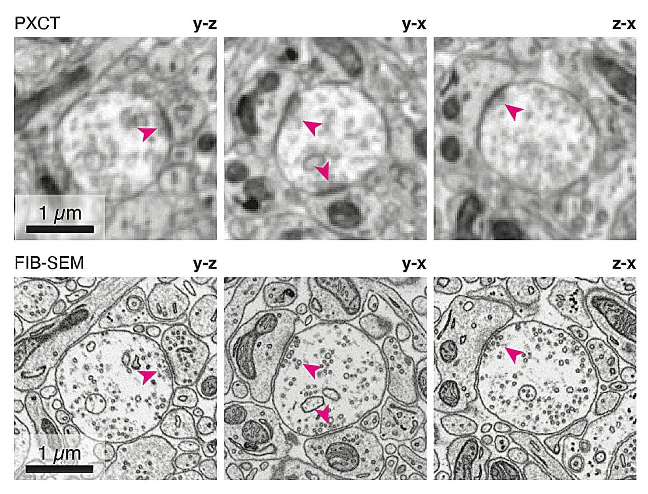
An international team of researchers led by the Francis Crick Institute, working with the Paul Scherrer Institute, has developed a new imaging protocol to capture mouse brain cell connections in precise detail. In work published in Nature Methods, they combined the use of X-rays with radiation-resistant materials sourced from the aerospace industry.
The images acquired using this technique allowed the team to see how nerve cells connect in the mouse brain, without needing to thinly slice biological tissue samples.
Volume electron microscopy (volume EM) has been the gold standard for imaging how nerve cells connect as ‘“circuitry” inside the brain. It has paved the way for scientists to create maps called connectomes, of entire brains, first in fruit fly larvae and then the adult fruit fly. This imaging involves cutting 10s of nm thin slices (tens of thousands per mm of tissue), imaging each slice and then building the images back into their 3D structure.
Every day, our brains transform quick impressions, flashes of inspiration, and painful moments into enduring memories that underpin our sense of self and inform how we navigate the world. But how does the brain decide which bits of information are worth keeping—and how long to hold on?
Now, new research demonstrates that long-term memory is formed by a cascade of molecular “timers” unfolding across brain regions. With a virtual reality-based behavioral model in mice, the scientists discovered that long-term memory is orchestrated by key regulators that either promote memories into progressively more lasting forms or demote them until they are forgotten.
The findings, published in Nature, highlight the roles of multiple brain regions in gradually reorganizing memories into more enduring forms, with gates along the way to assess salience and promote durability.