A new drug candidate shows promise by reversing cognitive decline in advanced Alzheimer’s disease by restoring brain balance in an animal study.

The 2026 Timeline: AGI Arrival, Safety Concerns, Robotaxi Fleets & Hyperscaler Timelines ## The rapid advancement of AI and related technologies is expected to bring about a transformative turning point in human history by 2026, making traditional measures of economic growth, such as GDP, obsolete and requiring new metrics to track progress ## ## Questions to inspire discussion.
Measuring and Defining AGI
🤖 Q: How should we rigorously define and measure AGI capabilities? A: Use benchmarks to quantify specific capabilities rather than debating terminology, enabling clear communication about what AGI can actually do across multiple domains like marine biology, accounting, and art simultaneously.
🧠 Q: What makes AGI fundamentally different from human intelligence? A: AGI represents a complementary, orthogonal form of intelligence to human intelligence, not replicative, with potential to find cross-domain insights by combining expertise across fields humans typically can’t master simultaneously.
📊 Q: How can we measure AI self-awareness and moral status? A: Apply personhood benchmarks that quantify AI models’ self-awareness and requirements for moral treatment, with Opus 4.5 currently being state-of-the-art on these metrics for rigorous comparison across models.
AI Capabilities and Risks.
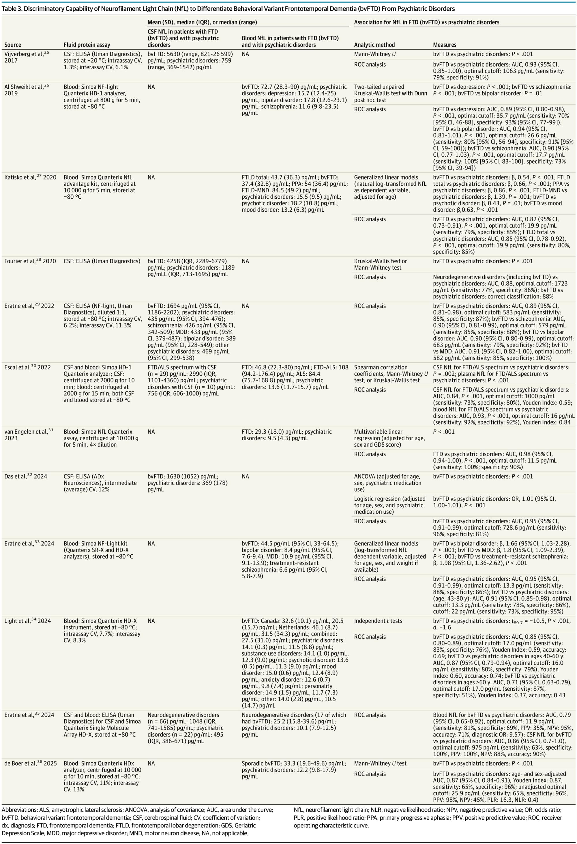
Neurofilament light chain levels in cerebrospinal fluid and blood are higher in patients with behavioral variant frontotemporal dementia than psychiatric disorders, suggesting its potential as a biomarker to aid in differentiating these conditions.
This systematic review aimed to determine whether elevated CSF or blood NfL can aid clinicians in differentiating bvFTD from psychiatric disorders. Across 12 studies reviewed, CSF and blood NfL levels were consistently significantly higher in patients with bvFTD than patients with psychiatric disorders at the group level. Furthermore, CSF and blood NfL demonstrated reasonable sensitivity and specificity to differentiate bvFTD from psychiatric disorders,38 though classification accuracy varied somewhat by study. While some of the AUCs for NfL differentiating bvFTD from psychiatric disorders had wide confidence intervals, these findings suggest a possible role for NfL in diagnostic clarification of patients with neuropsychiatric presentations. Misdiagnosing bvFTD as a psychiatric disorder may delay patients with bvFTD from early access to clinical trials when treatments may be more effective. Conversely, misdiagnosing a psychiatric disorder as bvFTD may worsen quality of life and delay treatment of ameliorable conditions.39-41
The existing literature has important limitations. Most studies were case control in design since they enrolled patients meeting diagnostic criteria for bvFTD or psychiatric disorders and then examined the association of NfL with these diagnoses. While these studies are essential for determining construct validity, they are not representative of the real-world clinical conditions by which NfL would be used. Also, such methods risk biased interpretations of the relationship of NfL with these conditions. Further studies are needed that prospectively recruit patients with neuropsychiatric symptoms and obtain NfL using clinically available assays to aid in real-time differential diagnosis.
An additional limitation of the reviewed studies is the selection and heterogeneity of psychiatric disorders. Several of the reviewed studies included patients with primary substance use, adjustment, or functional neurological disorder diagnoses.27, 31, 32,34, 35 Mood disorders, schizophrenia, and OCD may have the greatest symptom overlap with bvFTD.42 However, only 5 studies reported enrolling a total of 9 patients with OCD,25, 29, 31, 35, 36 a condition with an estimated OR of onset of 4.9 among individuals aged 45 to 59 years,43 suggesting a need for studies that include more patients with psychiatric disorders more likely to mimic core features of bvFTD despite absence of suspected neurodegenerative pathology. Moreover, FTD phenocopy syndromes (phFTD), clinical features that mimic FTD of mid-to late-life onset but do not progress to dementia, are understudied.44 Only 1 study in this review included a total of 2 patients with phFTD.

Periodontitis is widespread and can have serious consequences for overall health. Researchers at Fraunhofer have identified a substance that selectively inhibits only those bacteria that cause periodontitis, thereby preserving the natural balance of the oral microbiome. This technology has been further developed and commercialized as a range of oral care products by the spin-off company PerioTrap.
The oral microbiome is home to more than 700 different bacterial species, of which only a few can cause periodontitis. These adhere to dental plaque, particularly along the gum line, where they trigger inflammation (gingivitis). This can potentially lead to chronic periodontitis, which does more than just cause receding gums and loose teeth. If these bacteria enter the bloodstream, they can also contribute to the development of diabetes, rheumatic disease, arthritis, cardiovascular disease, chronic inflammatory bowel disease and even Alzheimer’s disease.
Pathogenic bacteria are killed by conventional oral care products such as alcohol-based mouthwashes and products containing the antiseptic chlorhexidine, but these also eliminate beneficial microorganisms. When the oral microbiome re-establishes itself after treatment, pathogenic bacteria such as Porphyromonas gingivalis gain an early advantage because they proliferate particularly well in inflamed gum tissue. Beneficial bacteria grow more slowly, and the oral microbiome quickly shifts back from its natural balance into dysbiosis, allowing the disease to recur.
But there is nothing in biology yet found that indicates the inevitability of death. This suggests to me that it is not at all inevitable, and that it is only a matter of time before the biologists discover what it Читать
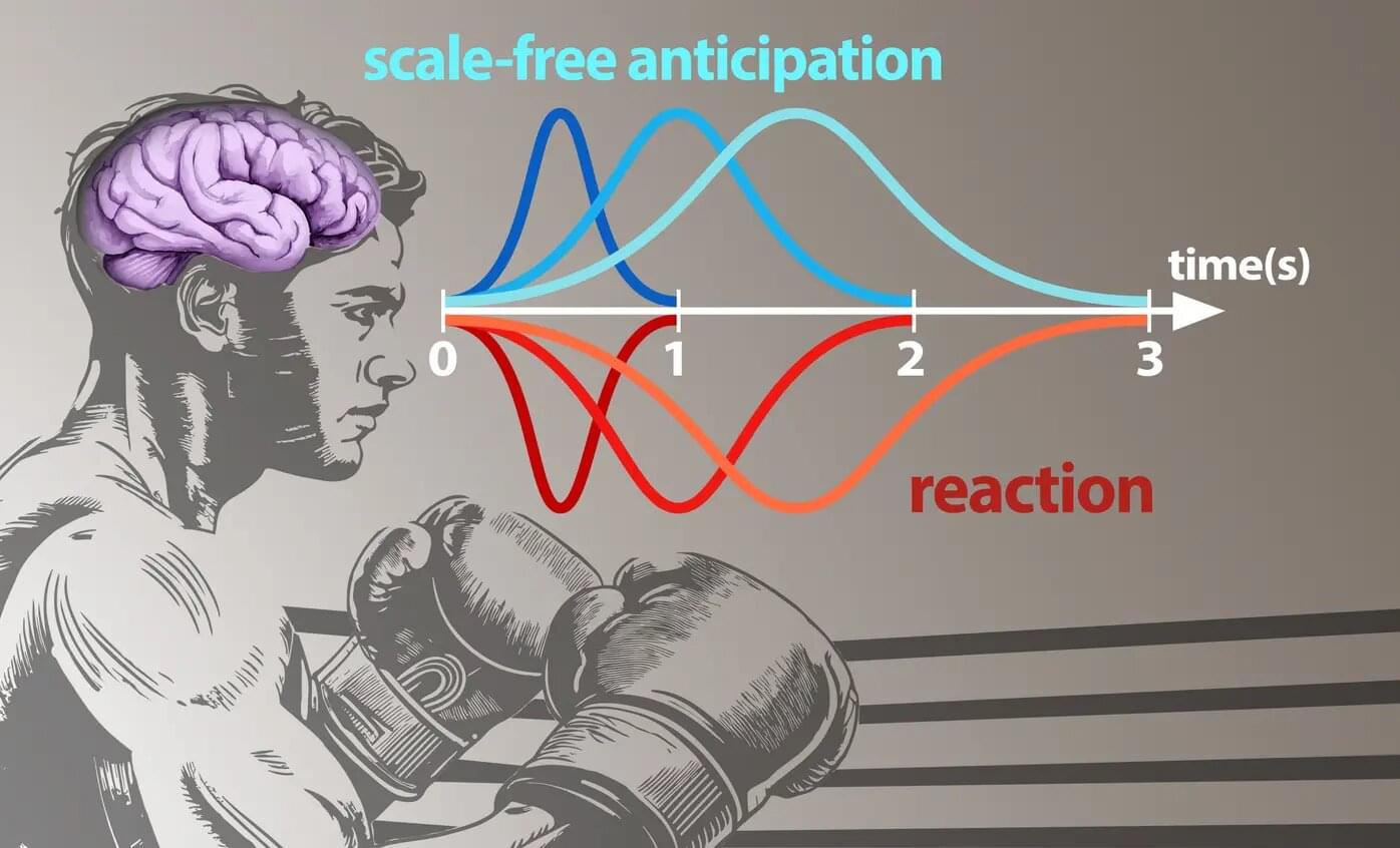
Humans respond to environments that change at many different speeds. A video game player, for example, reacts to on-screen events unfolding within hundreds of milliseconds or over several seconds. A boxer anticipates an opponent’s moves—even when their timing differs from that of previous opponents. In each case, the brain predicts when events occur, prepares for what comes next and flexibly adapts to the demands of the situation.
A study by neuroscientists from the Ernst Strüngmann Institute of the Max Planck Society, Goethe University Frankfurt, the Max Planck Institute for Empirical Aesthetics, and New York University, explains how the human brain predicts the timing of future events.
The research, published in the journal Proceedings of the National Academy of Sciences, shows that the brain continuously estimates how likely something is to happen within the next three seconds—and uses this estimate to prepare fast and accurate reactions.
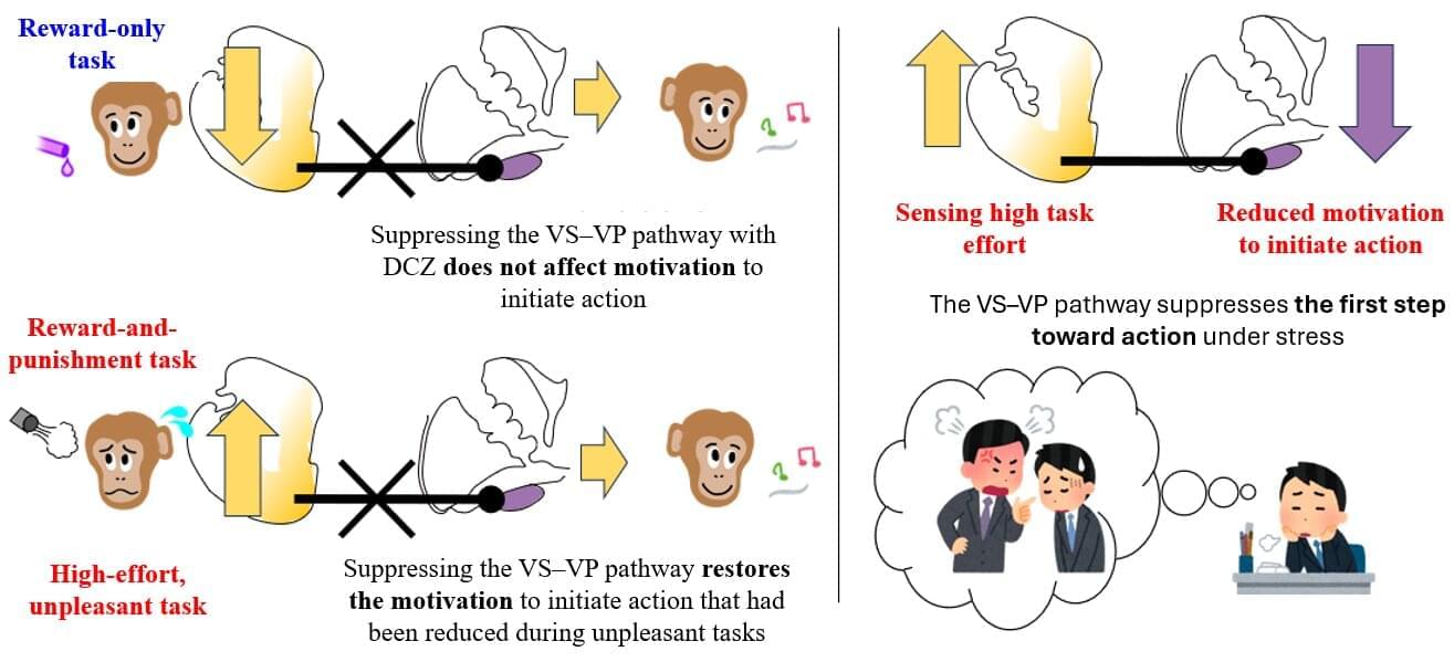
Most of us know the feeling: maybe it is making a difficult phone call, starting a report you fear will be criticized, or preparing a presentation that’s stressful just to think about. You understand what needs to be done, yet taking that very first step feels surprisingly hard.
When this difficulty becomes severe, it is known medically as avolition. People with avolition are not lazy or unaware: they know what they need to do, but their brain seems unable to push the “go” button.
Avolition is commonly seen in conditions such as depression, schizophrenia, and Parkinson’s disease, and it seriously disrupts a person’s ability to manage daily life and maintain social functions.
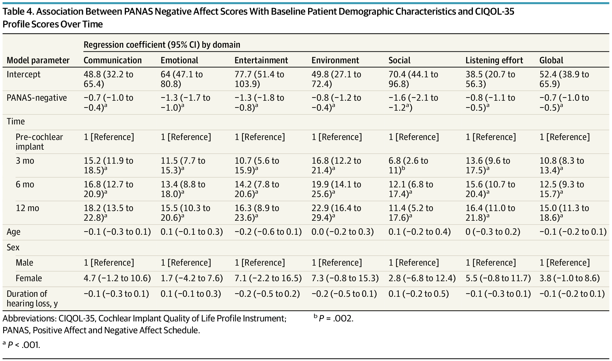
In adults receiving cochlear implants, gains in positive affect and reductions in negative affect corresponded with improvements in quality-of-life scores across listening, communication, and participation domains. Strongest statistical associations were observed in social and emotional CIQOL areas, but effect sizes were small.
Importance The use of patient-reported outcome measures to assess outcomes in adults who use cochlear implants has increased, as highlighted by the inclusion of the Cochlear Implant Quality of Life (CIQOL) instruments in the Minimal Speech Testing Battery, version 3. However, the self-reported nature of these instruments raises questions regarding how psychosocial characteristics impact responses.
Objective To assess whether affect and CIQOL domain scores change over time and whether affect is associated with CIQOL domain scores.
Design, Setting, and Participants Prospective longitudinal cohort study in adult cochlear implant candidates (aged 18–89 years) meeting indications for cochlear implantation based on bilateral moderate to profound hearing loss with aided sentence recognition scores 60% or less between September 19, 2019, and October 8, 2021, in a single tertiary otolaryngology referral center. Patients receiving a second cochlear implant and those without Montreal Cognitive Assessment scores were excluded. Follow-up duration was 1 year. Data analysis was performed between October 15, 2023, and August 5, 2025.