Parkinson’s disease (PD) is a debilitating and progressive neurodegenerative disorder caused by the loss of dopamine-producing neurons in the substantia nigra, a brain region essential for motor control. Clinically, it is marked by tremor, rigidity, bradykinesia and postural instability, symptoms that progressively erode independence and quality of life.
PD affects millions of people worldwide, including nearly one million individuals in the United States, making it one of the fastest-growing neurological disorders. In the U.S. alone, the disease imposes a profound health care and socioeconomic burden, with annual costs reaching tens of billions of dollars due to medical care, lost productivity and long-term disability.
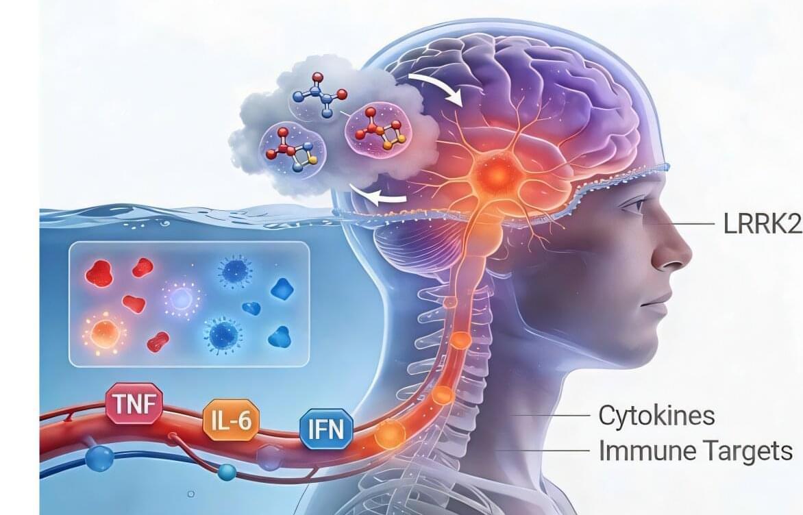
While environmental factors contribute to disease risk, genetic drivers are increasingly recognized, with mutations in the leucine-rich repeat kinase 2 (LRRK2) gene representing one of the most common causes of both familial and sporadic PD. Understanding how LRRK2 mutations drive disease is therefore central to developing therapies that go beyond symptoms control.