Nice.
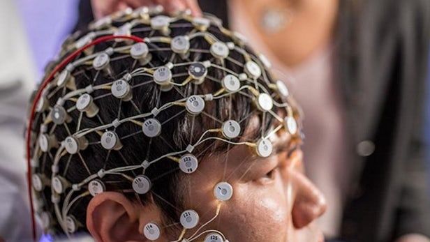
Rugby teams and ambulance staff are the target for portable brain scanner.
MINNEAPOLIS — Scanning a premature infant’s brain shortly after birth to map the location and volume of lesions, small areas of injury in the brain’s white matter, may help doctors better predict whether the baby will have disabilities later, according to a new study published in the January 18, 2017, online issue of Neurology ®, the medical journal of the American Academy of Neurology.
According to the Centers for Disease Control and Prevention, one in 10 babies is born prematurely in the United States.
Lack of oxygen to the brain is the most common form of brain injury in premature infants, resulting in damage to the white matter. White matter contains nerve fibers that maintain contact between various parts of the brain. Damage to white matter can interfere with communication in the brain and the signals it sends to other parts of the body.
Biological engineers at Harvard University’s Wyss Institute for Biologically Inspired Engineering have invented a microchip that can be lined with living human cells in order to revolutionise medicine, particularly relating to drug testing, disease modelling and personalised medicine.
The ‘human organs-on-chip’ is a microchip made from a clear flexible polymer that contains hollow microfluidic channels that are lined with living human cells, together with an interface that lines the interior surface of blood vessels and lymphatic vessels, known as an endothelium.
The idea is that the microchip can emulate the microarchitecture and functions of multiple human organs such as the lungs, kidneys, skin, bone marrow, intestines and blood-brain barrier. And if you were able to do this, you could then test out drugs and study how diseases affect the body without having to endanger human patients, or waste precious organs needed for transplants.
Summary: Researchers report an anti malaria drug has helped improve length and quality of life for a 26 year old brain cancer patient.
Source: University of Colorado.
After her brain cancer became resistant to chemotherapy and then to targeted treatments, 26-year-old Lisa Rosendahl’s doctors gave her only a few months to live. Now a paper published January 17 in the journal eLife describes a new drug combination that has stabilized Rosendahl’s disease and increased both the quantity and quality of her life: Adding the anti-malaria drug chloroquine to her treatment stopped an essential process that Rosendahl’s cancer cells had been using to resist therapy, re-sensitizing her cancer to the targeted treatment that had previously stopped working. Along with Rosendahl, two other brain cancer patients were treated with the combination and both showed similar, dramatic improvement.
Pretty wild.
Researchers of the German Center for Neurodegenerative Diseases (DZNE) have found that “alpha-synuclein,” a protein involved in a series of neurological disorders including Parkinson’s disease, is capable of travelling from brain to stomach and that it does so following a specific pathway. Donato Di Monte and co-workers report on this in the journal Acta Neuropathologica. Their study, carried out in rats, sheds new light on pathological processes that could underlie disease progression in humans.
Alpha-synuclein occurs naturally in the nervous system, where it plays an important role in synaptic function. However, in Parkinson’s disease, dementia with Lewy bodies and other neurodegenerative diseases termed “synucleinopathies,” this protein is accumulated within neurons, forming pathological aggregates. Distinct areas of the brain become progressively affected by this condition. The specific mechanisms and pathways involved in this widespread distribution of alpha-synuclein pathology remain to be fully elucidated. Clinical and experimental evidence suggests however that alpha-synuclein — or abnormal forms of it — could “jump” from one neuron to another and thus spread between anatomically interconnected regions.
Alpha-synuclein lesions have also been observed within neurons of the peripheral nervous system, such as those in the gastric wall. In some Parkinson’s patients, these lesions were detected at early disease stages. “Based on these intriguing observations, it has been hypothesized that the pathological process underlying Parkinson’s disease may actually start in the gastrointestinal tract and then move toward the brain,” Professor Di Monte says. “Our present approach was to look at this long-distance transmission of alpha-synuclein from the opposite perspective, investigating the possibility that alpha-synuclein may travel from the brain to the gut.”
Hmmm; you have a 2nd brain inside your stomach. At least this new research is pointing to that.
Gut feelings influence your mood and well-being.
Nice.
In research that could one day lead to advances against neurodegenerative diseases like Alzheimer’s and Parkinson’s, University of Michigan engineering researchers have demonstrated a technique for precisely measuring the properties of individual protein molecules floating in a liquid.
Proteins are essential to the function of every cell. Measuring their properties in blood and other body fluids could unlock valuable information, as the molecules are a vital building block in the body. The body manufactures them in a variety of complex shapes that can transmit messages between cells, carry oxygen and perform other important functions.
Sometimes, however, proteins don’t form properly. Scientists believe that some types of these misshapen proteins, called amyloids, can clump together into masses in the brain. The sticky tangles block normal cell function, leading to brain cell degeneration and disease.
But the processes of how amyloids form and clump together are not well understood. This is due in part to the fact that there’s currently not a good way to study them. Researchers say current methods are expensive, time-consuming and difficult to interpret, and can only provide a broad picture of the overall level of amyloids in a patient’s system.
Exercise is one of the best ways to slow down aging and its free too!
(Boston) — Older adults who experience good cardiac fitness may be also keeping their brains in good shape as well.
In what is believed to be the first study of its kind, older adults who scored high on cardiorespiratory fitness (CRF) tests performed better on memory tasks than those who had low CRF. Further, the more fit older adults were, the more active their brain was during learning. These findings appear in the journal Cortex. Difficulty remembering new information represents one of the most common complaints in aging and decreased memory performance is one of the hallmark impairments in Alzheimer’s disease.
Healthy young (18−31 years) and older adults (55−74 years) with a wide range of fitness levels walked and jogged on a treadmill while researchers assessed their cardiorespiratory fitness by measuring the ratio of inhaled and exhaled oxygen and carbon dioxide. These participants also underwent MRI scans which collected images of their brain while they learned and remembered names that were associated with pictures of unfamiliar faces.
Many who worked closely with me at Microsoft use to say I had a Crystal ball and was psychic; maybe I have met my match for a career — LOL.
A number of job adverts suggest that Facebook is taking social networking to a different level of science fiction.
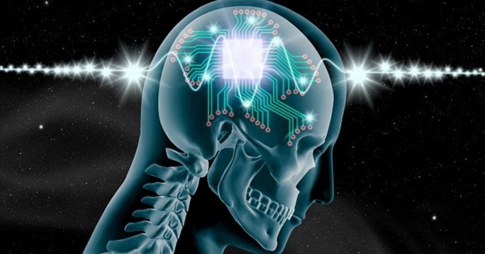
The social networking giant has advertised for a Haptics Engineer, a Neural Imaging Engineer, a Signal Processing Engineer and a Brain-Computer Interface (BCI) Engineer – leading people to think Facebook is working on mind reading technology.
This is supported by each of the job descriptions. The roles are based in Facebook’s elusive ‘Building 8’ in California, where the team will “apply DARPA-style breakthrough development at the intersection of ambitious science and product development.”
But, the larger question remains as to how these individual dendrites and neuron outputs are used by the circuit and the brain as a whole. These findings are considerably different than sequences needing a group of neurons working in order and in a circuit. Even more unusual is the fact that (even young childrens’) brains are able to analyze and respond to information that is, in fact, so complex that the most advanced super computers cannot. Can individual cells do this as well?
Another new set of research shows that in a monkey brain, these responses of individual neurons are correlated somewhat with the final decision of the animal. This research used very limited visual information and showed that the final decisions of the animal using billions of neurons was perhaps relevant even to this small amount of information input given to individual cells.
It could be that the local neuron responded to the decision that was made by the larger circuits and brain. But, it doesn’t answer the question as to how the individual neuron relates to the brain.