The octopus essentially has many brains and can edit its own dna this is probably the closest invebrate as close to an alien species that we have on earth because their biology is so exotic.
Category: neuroscience – Page 1,084

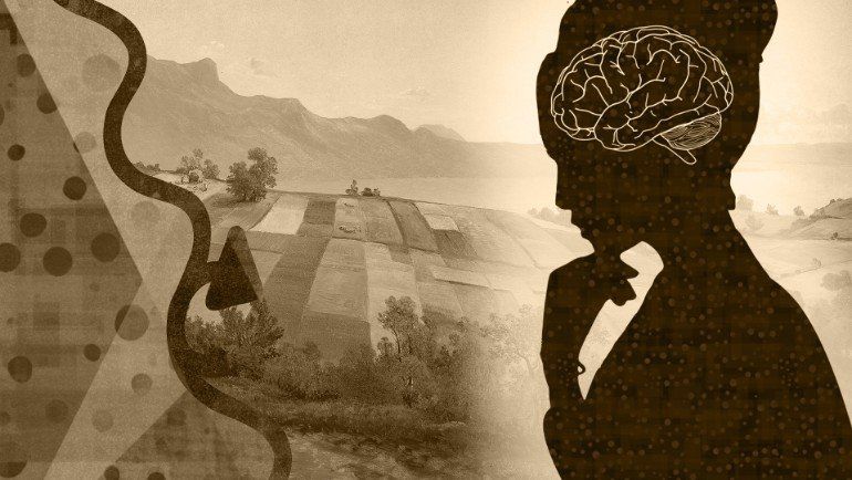
This New AI-Driven Quiz Predicts Your Psychological Age
A questionnaire developed by XPRIZE and Deep Longevity claims to be able to accurately predict your psychological age by using artificial intelligence (AI) to analyze your answers, which theoretically should fall close to your chronological age. The questionnaire is developed from a study published in Aging that used AI in an attempt to identify key hallmarks of psychological aging and the top risk factors that affect mortality.
While the questionnaire seems like a fun insight into whether you’re an old soul or a young gun at heart, there is an important scientific basis for an age-predicting AI. The newly developed technology represents the first AI aimed solely at using psychological aspects to predict age. The researchers hope it can contribute to understanding the role psychological clocks play in overall aging, as well as improving mental health and the feeling of youth.
“For the first time, AI can predict human psychological and subjective age and help identify the possible interventions that can be applied in order to help people feel and behave younger,” said Alex Zhavoronkov, PhD, founder and CLO of Deep Longevity and co-author of the study, in a statement.
TIMELAPSE OF FUTURE TECH: From 2022 — 4000+
The journey to see future technology starts in 2022, when Elon Musk and SpaceX send the first Starship to Mars — beginning the preparations for the arrival of the first human explorers.
We see the evolution of space exploration, from NASA’s Artemis mission, humans landing on Mars, and the interplanetary internet system going online. To the launch of the Starshot Alpha Centauri program, and quantum computers designing plants that can survive on Mars.
On Earth, tech evolves with quantum computers and Neaulink chips. People begin living with bio-printed organs. Humans record every part of lives from birth. And inner speech recording becomes possible.
And what about predictions further out into the future, when humans become level 2 and level 3 civilizations. When NASA’s warp drive goes live, and Mars declares independence from Earth. Will there be Dyson structures built around stars to capture their energy. Will they help power computers that can take human consciousness and download it into a quantum computer core. Allowing humanity to travel further out into space.
Quotes about the future from: Arthur C. Clarke, Stephen Hawking, Albert Einstein, and Elon Musk.
Additional footage sourced from: NASA, SpaceX.
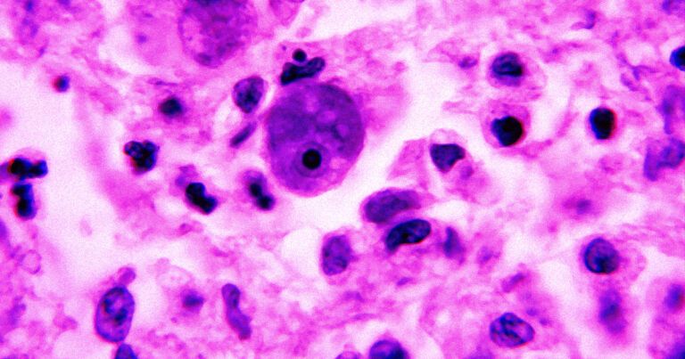
Brain-Eating Amoeba Is Spreading in United States, Scientists Say
The single-celled organism behind the infections is usually found in warm bodies of freshwater, including lakes and rivers. Once a person is infected — an exceedingly rare occurrence usually resulting from swimming or diving in infected waters — the amoeba travels from the nose into the brain.
Once there, the organism can kick off a nasty brain condition called primary amebic meningoencephalitis (PAM). And yep, according to the CDC, PAM is “usually fatal.”
The good news: there have only been 34 infections reported in the US in the last ten years, according to CDC data.
Cellular Connections Found Between Nervous and Immune Systems
Summary: Researchers have identified a direct cellular interaction between the nervous system and the immune system. Pain sensing neurons around the lymph nodes can modulate lymph node activity.
Source: Broad Institute.
The nervous and immune systems have long been thought to be separate entities in the body, but new research has uncovered a direct cellular interaction between the two. Scientists from Harvard Medical School, the Broad Institute of MIT and Harvard, MIT, and the Ragon Institute of MGH, MIT and Harvard have found that pain-sensing neurons surround lymph nodes in mice, and can modulate the activity of these small organs, which are key parts of the immune system.
Protein Involved in Removing Alzheimer’s Buildup Linked to Circadian Rhythm
Summary: YKL-40 may be a key player in circadian rhythm disruptions associated with Alzheimer’s disease.
Source: WUSTL
Fractured sleep, daytime sleepiness and other signs of disturbance in one’s circadian rhythm are common complaints of people with Alzheimer’s disease, and the problems only get worse as the disease progresses. But the reason for the link between Alzheimer’s and circadian dysfunction is not well understood.

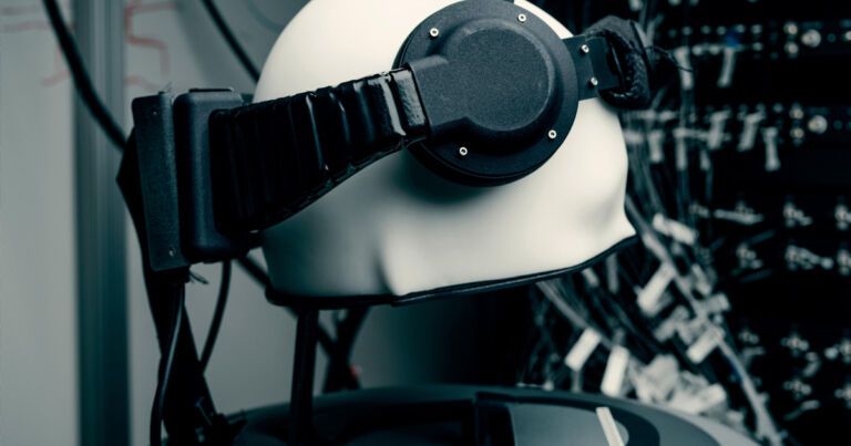
Leaked Meeting: Facebook Working on Device to Read Your Brain
Pre-Emptive Defense
All the same, Schroepfer seemed to acknowledge that getting people to actually use the tech might be a hard sell, according to the leaked meeting audio acquired by BuzzFeed News.
“We have to build responsibly to earn trust and the right to continue to grow,” Schroepfer said. “It’s imperative that we get this right so that people around the world get all these amazing technologies… without experiencing the downsides.”

Raccoon intelligence at the borderlands of science
All hail the powerful One climbed my wood structure that went straight up then went to the roof o.o. Also their hands make them like chimps.
How does intelligence ofs compare with other species? That was a topic of heated debate between 1905 and 1915 within the then-nascent field of comparative psychology.
In 1907, psychologist Lawrence W. Cole, who had established a colony ofs at the University of Oklahoma, and Herbert Burnham Davis, a doctoral student at Clark University, each published the results of nearly identical experiments on the processes of learning, association and memory ins. They relied on E.L. Thorndike’s puzzle-box methodology, which involved placing animals in wooden crates from which the animal had to escape by opening the latch or sequence of latches. They observed the number of trials required for successful completion and the extent to which the animal retained the ability to solve the same problem more quickly when confronted again with it. Using this method, they sought what Davis called “a tolerable basis” for ranking the intelligence ofs on the phylogenetic scale of evolutionary development. They independently concluded thats bested the abilities of cats and dogs, most closely approximating the mental attributes of monkeys.
Raccoons had attracted interest because they flourished, rather than receded, in the face of human expansion. Over the centuries, people had hunteds for food and fur, decried them as agricultural pests and urban bandits, and kept them as household pets. This latter role brought the species to psychologists’ attention. Cole reported that he got the idea to work withs from observing the behavior of a pet kept at a local market. At the time, most animal experiments being conducted occurred on the borderlands of academic research, nature study and domestic life. Scientists such as Charles Darwin, William James and James Mark Baldwin all developed psychological theories based upon observations of their own children and pets. Cole’ss, for example, lived simultaneously as research objects and amusing pets, a relationship that shaped how these experiments were presented to and perceived by the public. Despite Davis’s protests, a widely printed newspaper story depicted his puzzle-box experiments as an example of teaching “tricks” to one’s pets.
The DNA Regions in Our Brain That Contribute to Make Us Human
Summary: A new method identified a large set of gene regulatory regions in the brain, selected throughout human evolution.
Source: Swiss Institute of Bioinformatics.
With only 1% difference, the human and chimpanzee protein-coding genomes are remarkably similar. Understanding the biological features that make us human is part of a fascinating and intensely debated line of research. Researchers at the SIB Swiss Institute of Bioinformatics and the University of Lausanne have developed a new approach to pinpoint, for the first time, adaptive human-specific changes in the way genes are regulated in the brain.