Find out more about What are nanorobots or nanobots?, don’t miss it.
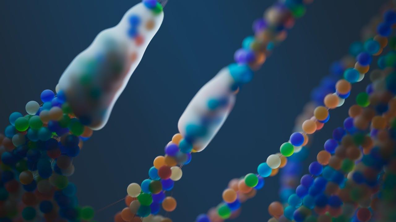


Apical periodontitis, a chronic and hard-to-treat dental infection, affects more than half of the population worldwide and is the leading cause of tooth loss. Root canal is the standard treatment, but existing approaches to treat the infection have many limitations that can cause complications, leading to treatment failure.
Now, researchers at the School of Dental Medicine, Perelman School of Medicine, and School of Engineering and Applied Sciences have identified a promising new therapeutic option that could potentially disrupt current treatments. The team of researchers is part of the Center for Innovation & Precision Dentistry, a joint research center between Penn Dental Medicine and Penn Engineering that leverages engineering and computational approaches to advance oral and craniofacial health care innovation.
In a paper published in the Journal of Clinical Investigation, they show that ferumoxytol, an FDA-approved iron oxide nanoparticle formulation, greatly reduces infection in patients diagnosed with apical periodontitis.
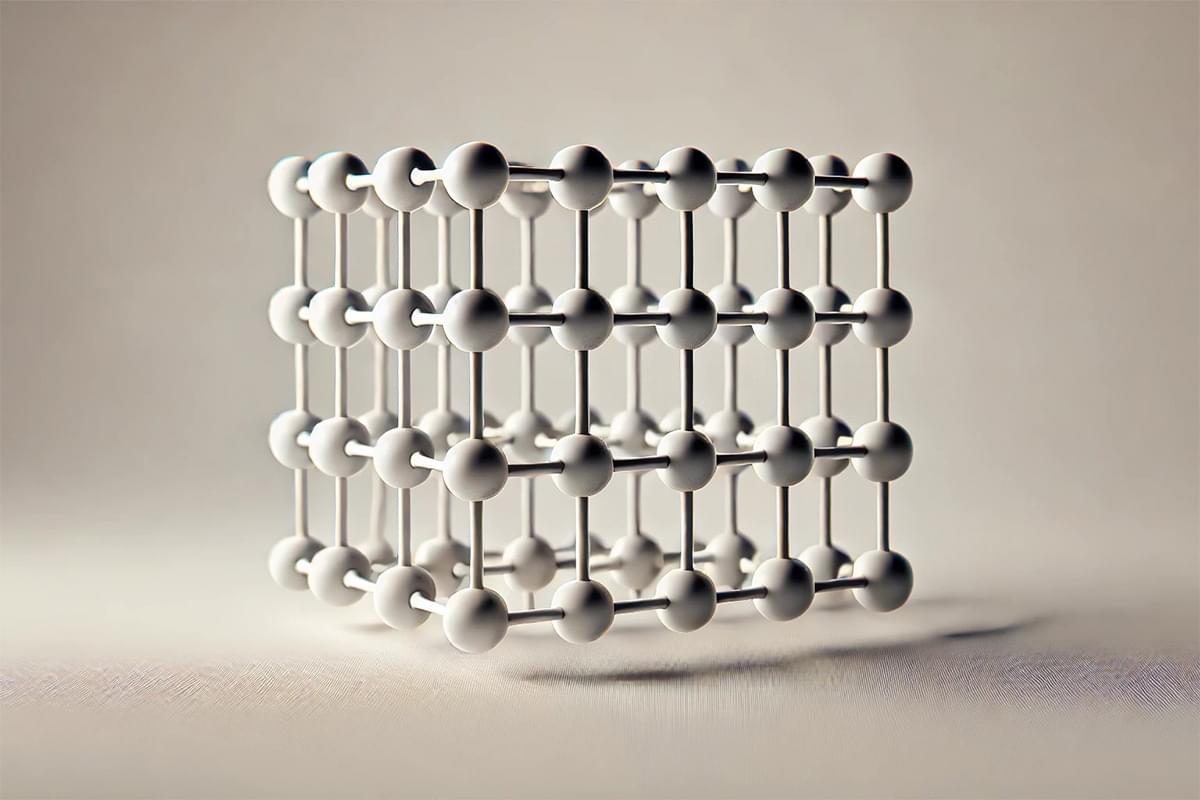
Using machine learning, a team of researchers in Canada has created ultrahigh-strength carbon nanolattices, resulting in a material that’s as strong as carbon steel, but only as dense as Styrofoam.
The team noted last month that it was the first time this branch of AI had been used to optimize nano-architected materials. University of Toronto’s Peter Serles, one of the authors of the paper describing this work in Advanced Materials, praised the approach, saying, “It didn’t just replicate successful geometries from the training data; it learned from what changes to the shapes worked and what didn’t, enabling it to predict entirely new lattice geometries.”
To quickly recap, nanomaterials are engineered by arranging atoms or molecules in precise patterns, much like constructing structures with extremely tiny LEGO blocks. These materials often exhibit unique properties due to their nanoscale dimensions.
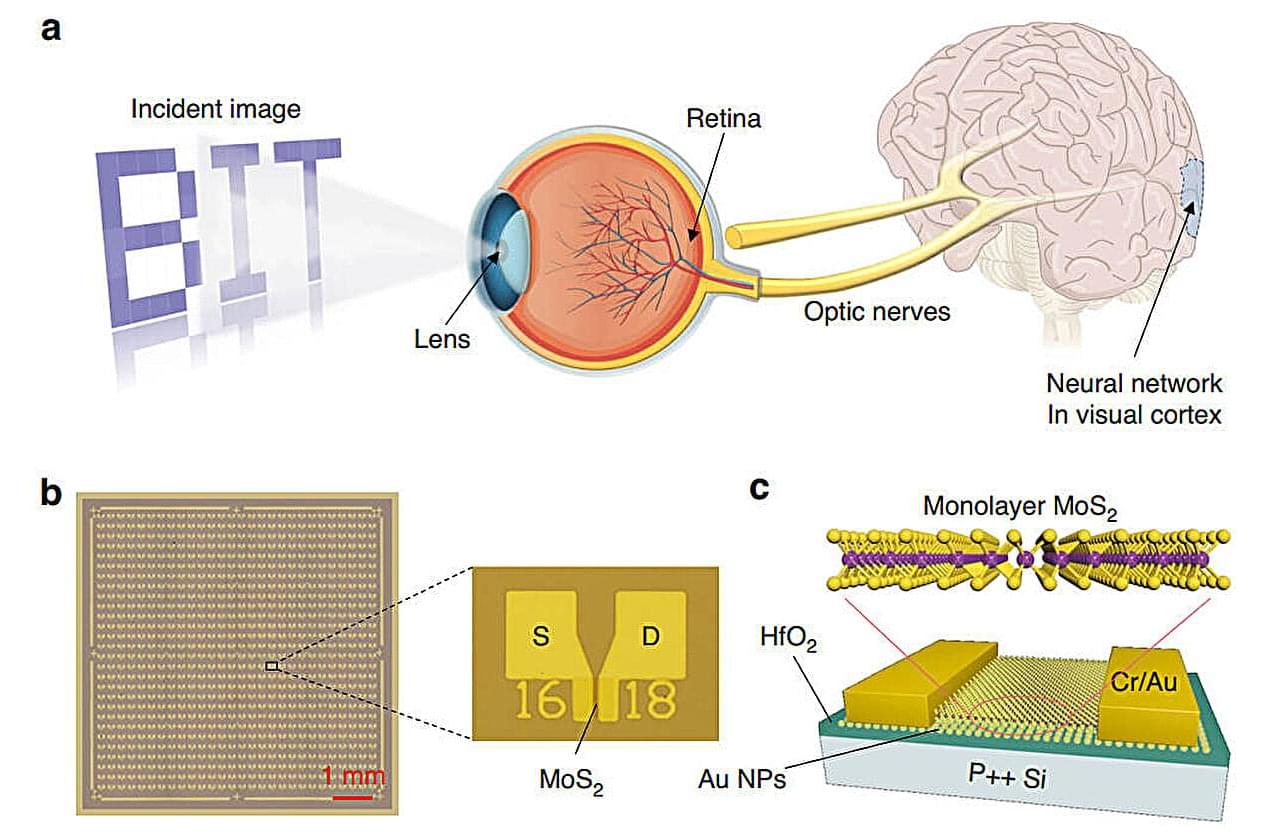
In a development for artificial intelligence, researchers have unveiled a synaptic device array that shows promise for enhancing artificial visual systems. This innovative array, measuring a compact 0.7 × 0.7 cm2, integrates the capabilities of sensing, memory, and processing to mimic the intricate functions of the human visual system.
Utilizing wafer-scale monolayer molybdenum disulfide (MoS2) and gold nanoparticles for enhanced electron capture, the array exhibits remarkable coordination between optical and electrical components. It is capable of both writing and erasing images and has achieved a 96.5% accuracy in digit recognition, marking a significant leap forward in the development of large-scale neuromorphic systems.
The human visual system processes complex visual data efficiently through an interconnected network that allows for parallel processing. However, current artificial vision systems face numerous challenges, including circuit complexity, high power consumption, and difficulties in miniaturization.

At the threshold of a century poised for unprecedented transformations, we find ourselves at a crossroads unlike any before. The convergence of humanity and technology is no longer a distant possibility; it has become a tangible reality that challenges our most fundamental conceptions of what it means to be human.
This article seeks to explore the implications of this new era, in which Artificial Intelligence (AI) emerges as a central player. Are we truly on the verge of a symbiotic fusion, or is the conflict between the natural and the artificial inevitable?
The prevailing discourse on AI oscillates between two extremes: on one hand, some view this technology as a powerful extension of human capabilities, capable of amplifying our creativity and efficiency. On the other, a more alarmist narrative predicts the decline of human significance in the face of relentless machine advancement. Yet, both perspectives seem overly simplistic when confronted with the intrinsic complexity of this phenomenon. Beyond the dichotomy of utopian optimism and apocalyptic pessimism, it is imperative to critically reflect on AI’s cultural, ethical, and philosophical impact on the social fabric, as well as the redefinition of human identity that this technological revolution demands.
Since the dawn of civilization, humans have sought to transcend their natural limitations through the creation of tools and technologies. From the wheel to the modern computer, every innovation has been seen as a means to overcome the physical and cognitive constraints imposed by biology. However, AI represents something profoundly different: for the first time, we are developing systems that not only execute predefined tasks but also learn, adapt, and, to some extent, think.
This transition should not be underestimated. While previous technologies were primarily instrumental—serving as controlled extensions of human will—AI introduces an element of autonomy that challenges the traditional relationship between subject and object. Machines are no longer merely passive tools; they are becoming active partners in the processes of creation and decision-making. This qualitative leap radically alters the balance of power between humans and machines, raising crucial questions about our position as the dominant species.
But what does it truly mean to “be human” in a world where the boundaries between mind and machine are blurring? Traditionally, humanity has been defined by attributes such as consciousness, emotion, creativity, and moral decision-making. Yet, as AI advances, these uniquely human traits are beginning to be replicated—albeit imperfectly—within algorithms. If a machine can imitate creativity or exhibit convincing emotional behavior, where does our uniqueness lie?
This challenge is not merely technical; it strikes at the core of our collective identity. Throughout history, humanity has constructed cultural and religious narratives that placed us at the center of the cosmos, distinguishing us from animals and the forces of nature. Today, that narrative is being contested by a new technological order that threatens to displace us from our self-imposed pedestal. It is not so much the fear of physical obsolescence that haunts our reflections but rather the anxiety of losing the sense of purpose and meaning derived from our uniqueness.
Despite these concerns, many AI advocates argue that the real opportunity lies in forging a symbiotic partnership between humans and machines. In this vision, technology is not a threat to humanity but an ally that enhances our capabilities. The underlying idea is that AI can take on repetitive or highly complex tasks, freeing humans to engage in activities that truly require creativity, intuition, and—most importantly—emotion.
Concrete examples of this approach can already be seen across various sectors. In medicine, AI-powered diagnostic systems can process vast amounts of clinical data in record time, allowing doctors to focus on more nuanced aspects of patient care. In the creative industry, AI-driven text and image generation software are being used as sources of inspiration, helping artists and writers explore new ideas and perspectives. In both cases, AI acts as a catalyst, amplifying human abilities rather than replacing them.
Furthermore, this collaboration could pave the way for innovative solutions in critical areas such as environmental sustainability, education, and social inclusion. For example, powerful neural networks can analyze global climate patterns, assisting scientists in predicting and mitigating natural disasters. Personalized algorithms can tailor educational content to the specific needs of each student, fostering more effective and inclusive learning. These applications suggest that AI, far from being a destructive force, can serve as a powerful instrument to address some of the greatest challenges of our time.
However, for this vision to become reality, a strategic approach is required—one that goes beyond mere technological implementation. It is crucial to ensure that AI is developed and deployed ethically, respecting fundamental human rights and promoting collective well-being. This involves regulating harmful practices, such as the misuse of personal data or the indiscriminate automation of jobs, as well as investing in training programs that prepare people for the new demands of the labor market.
While the prospect of symbiotic fusion is hopeful, we cannot ignore the inherent risks of AI’s rapid evolution. As these technologies become more sophisticated, so too does the potential for misuse and unforeseen consequences. One of the greatest dangers lies in the concentration of power in the hands of a few entities, whether they be governments, multinational corporations, or criminal organizations.
Recent history has already provided concerning examples of this phenomenon. The manipulation of public opinion through algorithm-driven social media, mass surveillance enabled by facial recognition systems, and the use of AI-controlled military drones illustrate how this technology can be wielded in ways that undermine societal interests.
Another critical risk in AI development is the so-called “alignment problem.” Even if a machine is programmed with good intentions, there is always the possibility that it misinterprets its instructions or prioritizes objectives that conflict with human values. This issue becomes particularly relevant in the context of autonomous systems that make decisions without direct human intervention. Imagine, for instance, a self-driving car forced to choose between saving its passenger or a pedestrian in an unavoidable collision. How should such decisions be made, and who bears responsibility for the outcome?
These uncertainties raise legitimate concerns about humanity’s ability to maintain control over increasingly advanced technologies. The very notion of scientific progress is called into question when we realize that accumulated knowledge can be used both for humanity’s benefit and its detriment. The nuclear arms race during the Cold War serves as a sobering reminder of what can happen when science escapes moral oversight.
Whether the future holds symbiotic fusion or inevitable conflict, one thing is clear: our understanding of human identity must adapt to the new realities imposed by AI. This adjustment will not be easy, as it requires confronting profound questions about free will, the nature of consciousness, and the essence of individuality.
One of the most pressing challenges is reconciling our increasing technological dependence with the preservation of human dignity. While AI can significantly enhance quality of life, there is a risk of reducing humans to mere consumers of automated services. Without a conscious effort to safeguard the emotional and spiritual dimensions of human experience, we may end up creating a society where efficiency outweighs empathy, and interpersonal interactions are replaced by cold, impersonal digital interfaces.
On the other hand, this very transformation offers a unique opportunity to rediscover and redefine what it means to be human. By delegating mechanical and routine tasks to machines, we can focus on activities that truly enrich our existence—art, philosophy, emotional relationships, and civic engagement. AI can serve as a mirror, compelling us to reflect on our values and aspirations, encouraging us to cultivate what is genuinely unique about the human condition.
Ultimately, the fate of our relationship with AI will depend on the choices we make today. We can choose to view it as an existential threat, resisting the inevitable changes it brings, or we can embrace the challenge of reinventing our collective identity in a post-humanist era. The latter, though more daring, offers the possibility of building a future where technology and humanity coexist in harmony, complementing each other.
To achieve this, we must adopt a holistic approach that integrates scientific, ethical, philosophical, and sociological perspectives. It also requires an open, inclusive dialogue involving all sectors of society—from researchers and entrepreneurs to policymakers and ordinary citizens. After all, AI is not merely a technical tool; it is an expression of our collective imagination, a reflection of our ambitions and fears.
As we gaze toward the horizon, we see a world full of uncertainties but also immense possibilities. The future is not predetermined; it will be shaped by the decisions we make today. What kind of social contract do we wish to establish with AI? Will it be one of domination or cooperation? The answer to this question will determine not only the trajectory of technology but the very essence of our existence as a species.
Now is the time to embrace our historical responsibility and embark on this journey with courage, wisdom, and an unwavering commitment to the values that make human life worth living.
__
Copyright © 2025, Henrique Jorge
[ This article was originally published in Portuguese in SAPO’s technology section at: https://tek.sapo.pt/opiniao/artigos/a-sinfonia-do-amanha-tit…exao-seria ]
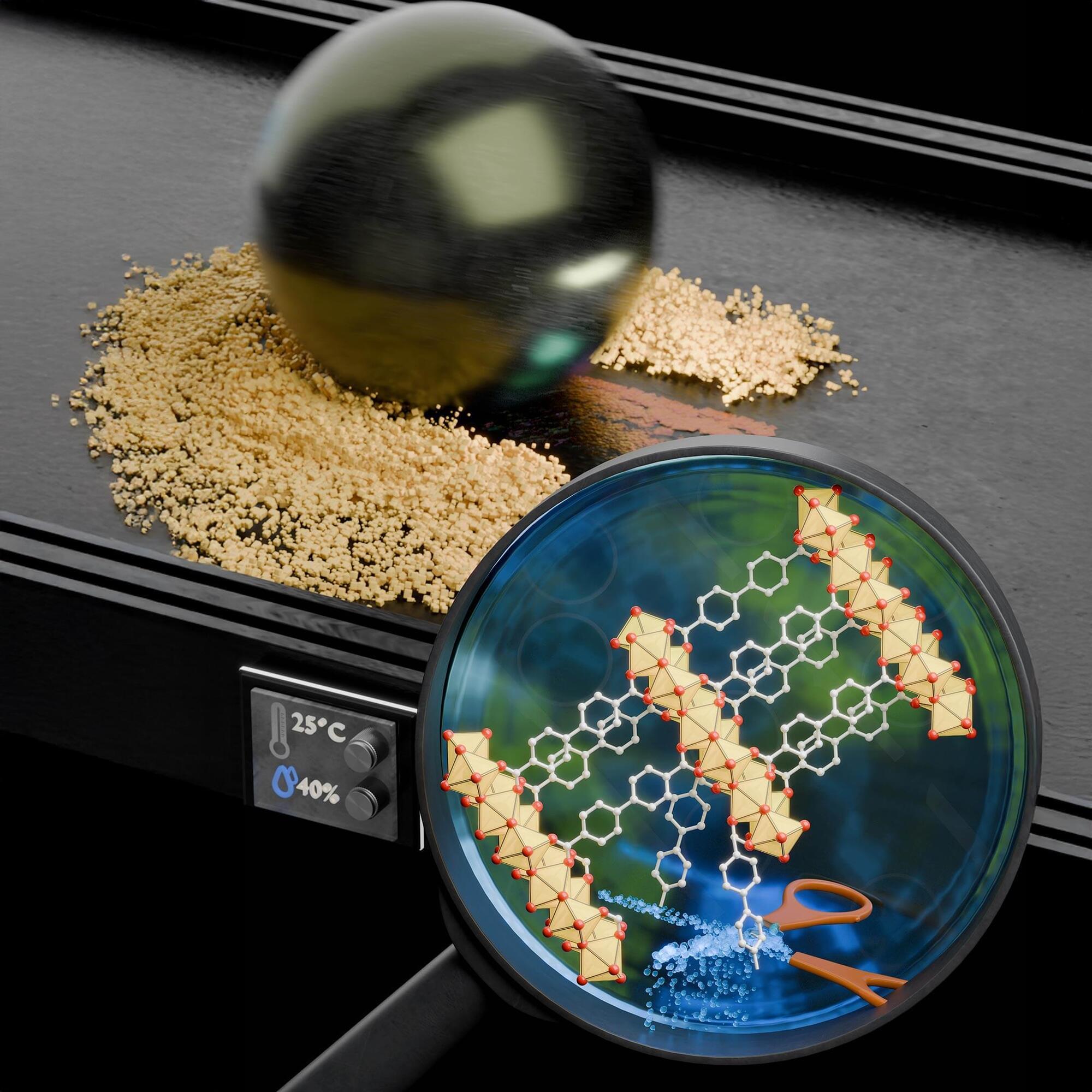
Researchers have developed COK-47, a solid lubricant that outperforms traditional options by leveraging water molecules to reduce friction.
This advanced material consists of ultra-thin titanium oxide sheets that create a low-friction tribofilm in humid conditions, making it highly durable and effective.
Revolutionizing Lubricants with Cutting-Edge Research.
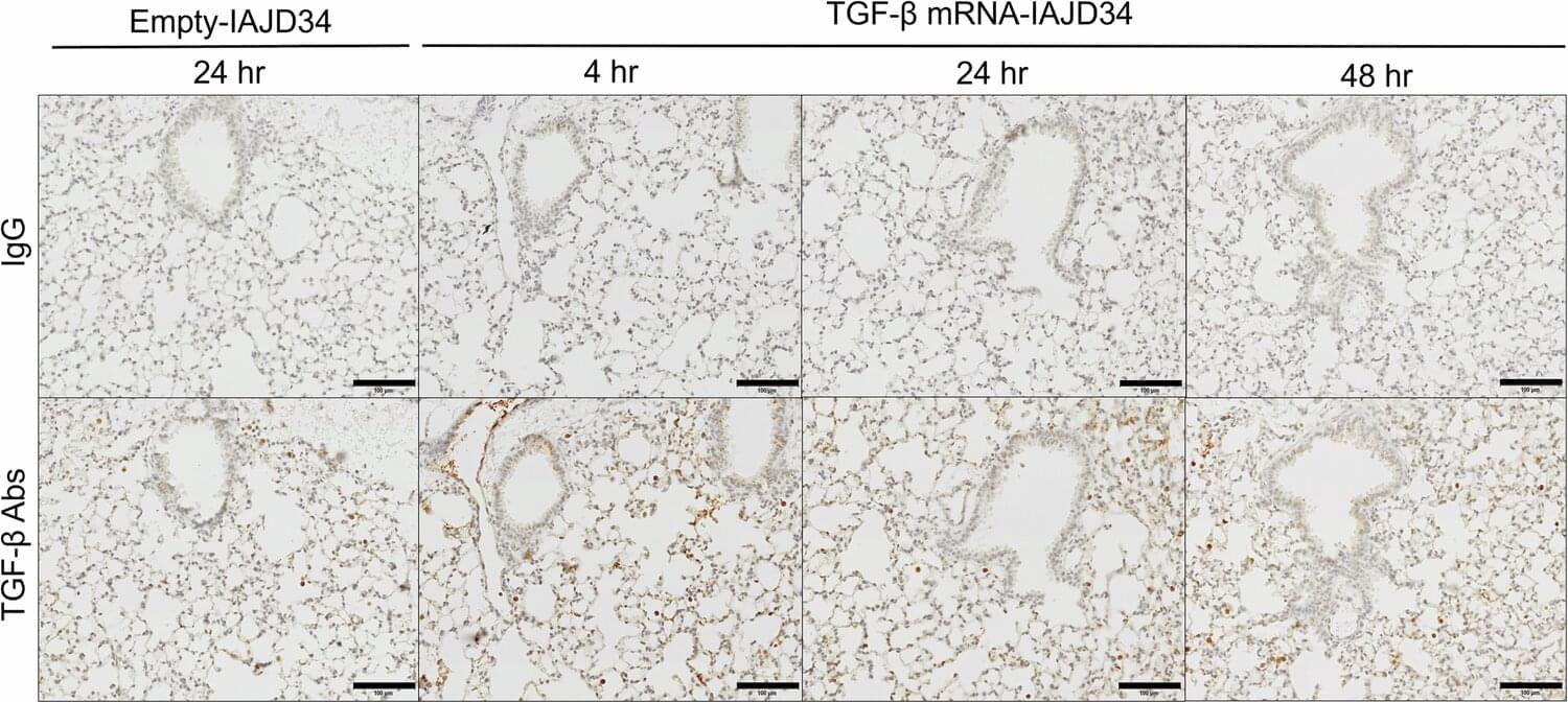
A combination of mRNA and a new lipid nanoparticle could help heal damaged lungs, according to new research from the Perelman School of Medicine at the University of Pennsylvania. Viruses, physical trauma, or other problems can have a serious impact on the lungs, and when the damage is in the lower regions, traditional treatments, like inhaled medication, might not work. The study, published in Nature Communications, provides a proof of concept for an injectable therapy.
“The lungs are hard-to-treat organs because both permanent and temporary damage often happen in the deeper regions where medication does not easily reach,” said study author Elena Atochina-Vasserman, MD, Ph.D., research assistant professor of Infectious Diseases at Penn and scientist at the Penn Institute for RNA Innovation. “Even drugs delivered intravenously are spread without specificity. That makes a targeted approach like ours especially valuable.”
Lung damage can result from a variety of causes ranging from physical accidents that cause inflammation of the lungs to respiratory viruses like COVID, flu, and RSV. Viruses alone can usher in an inflammatory response setting off a buildup of fluid in the airways, excess mucus, cell death, and damage to the lining of the lungs. Whether acute or chronic, weakened lungs can be life threatening. Respiratory diseases were the third leading cause of death worldwide even before the pandemic, according to previous research.
Scientists have created a hydrogel that strengthens bones in weeks. Bone density increased by 5X in a lab.
A groundbreaking injectable hydrogel may soon offer a faster, more effective treatment for osteoporosis, a condition that weakens bones and increases fracture risk.
Developed by researchers at EPFL in Switzerland and startup Flowbone, this new hydrogel, made from hyaluronic acid and hydroxyapatite nanoparticles, mimics bone’s natural minerals and strengthens fragile areas. In lab tests on rats, the treatment increased bone density by up to three times within weeks. When combined with the osteoporosis drug Zoledronate, bone density at the injection site increased nearly fivefold, potentially reducing the risk of fractures far more quickly than current medications.
While the hydrogel is not a permanent fix, researchers believe it could revolutionize osteoporosis management by complementing existing drug therapies and speeding up recovery. Given that osteoporosis affects millions worldwide—especially postmenopausal women—this breakthrough could significantly lower the risk of life-threatening fractures. The team now aims to secure regulatory approval and begin clinical trials, bringing this promising technology one step closer to real-world use. If successful, it could redefine how osteoporosis is treated, offering patients faster relief and stronger bones.
Managing osteoporotic patients at immediate fracture risk is challenging, in part due to the slow and localized effects of anti-osteoporotic drugs. Combining systemic anti-osteoporotic therapies with local bone augmentation techniques offers a promising strategy, but little is known about potential interactions. We hypothesized that integrating systemic treatments with local bone-strengthening biomaterials would have an additive effect on bone density and structure. This study investigated interactions and synergies between systemic therapies and injectable biomaterials, HA2 and HA2-ZOL, designed for local bone strengthening. HA2-ZOL incorporates Zoledronate, a bisphosphonate, to enhance anti-resorptive effects. These materials were tested in an in vivo rat model of osteoporosis using microCT and histology.
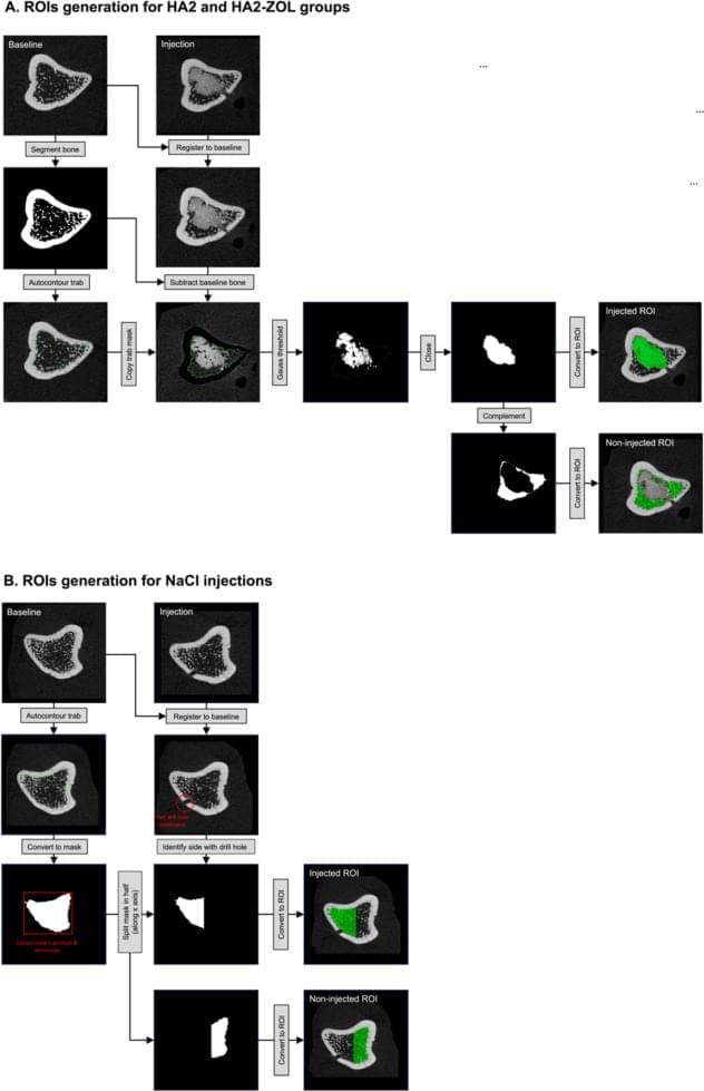
Thirty-six ovariectomized Wistar rats were treated systemically with vehicle (VEH), alendronate (ALN), or parathyroid hormone (PTH). One week later, their tibiae were randomly assigned to local treatment groups: HA2, HA2-ZOL, or NaCl control. Bilateral injections targeted metaphyseal trabecular bone, with microCT scans tracking changes over 8 weeks. Regions of interest (ROIs) were identified and analyzed for bone volume fraction (BV/TV), tissue mineral density (TMD), and trabecular morphology. Histological analyses were performed at week 8 to assess bone structure and mineral inclusions.

DGIST research teams have developed a self-powered sensor that uses motion and pressure to generate electricity and light simultaneously. This battery-free technology is expected to be used in various real-life applications, such as disaster rescue, sports, and wearable devices.
Triboelectric nanogenerators (TENG) and mechanoluminescence (ML) have attracted attention as green energy technologies that can generate electricity and light, respectively, without external power. However, researchers in previous studies mainly focused on the two technologies separately or simply combined them. Moreover, the power output stability of TENG and the insufficient luminous duration of ML materials have been major limitations for practical applications.
The research team has developed a system that generates electricity and light simultaneously using motion and pressure. They added light-emitting zinc sulfide-copper (ZnS: Cu) particles to a rubber-like material (polydimethylsiloxane [PDMS]) and designed a single electrode structure based on silver nanowires to obtain high efficiency. The developed device does not degrade in performance even after being repeatedly pressed more than 5,000 times, and it stably generates voltages of up to 60 V and a current of 395 nA.
As the fundamental flaw of today’s quantum computers, improving qubit stability remains the focus of much research in this field. One such stability attempt involves so-called topological quantum computing with the use of anyons, which are two-dimensional quasiparticles. Such an approach has been claimed by Microsoft in a recent paper in Nature. This comes a few years after an earlier claim by Microsoft for much the same feat, which was found to be based on faulty science and hence retracted.
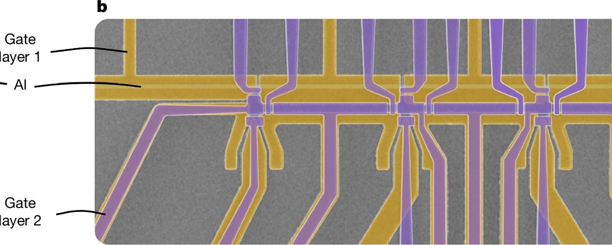
The claimed creation of anyons here involves Majorana fermions, which differ from the much more typical Dirac fermions. These Majorana fermions are bound with other such fermions as a Majorana zero mode (MZM), forming anyons that are intertwined (braided) to form what are in effect logic gates. In the Nature paper the Microsoft researchers demonstrate a superconducting indium-arsenide (InAs) nanowire-based device featuring a read-out circuit (quantum dot interferometer) with the capacitance of one of the quantum dots said to vary in a way that suggests that the nanowire device-under-test demonstrates the presence of MZMs at either end of the wire.
Microsoft has a dedicated website to their quantum computing efforts, though it remains essential to stress that this is not a confirmation until their research is replicated by independent researchers. If confirmed, MZMs could provide a way to create more reliable quantum computing circuitry that does not have to lean so heavily on error correction to get any usable output. Other, competing efforts here include such things as hybrid mechanical qubits and antimony-based qubits that should be more stable owing to their eight spin configurations.