In a major advancement in nanomedicine, Arizona State University (ASU) scientists, in collaboration with researchers from the National Center for Nanoscience and Technology (NCNST), of the Chinese Academy of Sciences, have successfully programmed nanorobots to shrink tumors by cutting off their blood supply.
Category: nanotechnology – Page 254
Researchers at the University of Chicago have developed light-activated nanowires that can stimulate neurons to fire when they are exposed to light. The researchers hope that the nanowires could help in understanding complex brain circuitry, and they may also be useful in treating brain disorders.
Optogenetics, which involves genetically modifying neurons so that they are sensitive to a light stimulus, has attracted a lot of attention as a research tool and potential therapeutic approach. However, some researchers have misgivings about optogenetics, as it involves inserting a gene into cells, potentially opening the door to unforeseen effects and possibly permanently altering treated cells.
In an effort to develop an alternative, a research team at the University of Chicago has devised a new modality that can enable light activation of neurons without the need for genetic modification. Their technique involves nanowires that are so small that if they were laid side-by-side, hundreds of them would fit on the edge of a sheet of paper. Although initially designed for use in solar cells, their small size also makes them well suited to interacting with cells.
Australian researchers from the ARC Centre of Excellence for Nanoscale BioPhotonics (CNBP) have developed a 3D printable ‘clip-on’ that can turn any smartphone into a fully functional microscope.
Reported in the research journal Scientific Reports, the smartphone microscope is powerful enough to visualise specimens as small as 1/200th of a millimetre, including microscopic organisms, animal and plant cells, blood cells, cell nuclei and more.
The clip-on technology is unique in that it requires no external power or light source to work yet offers high-powered microscopic performance in a robust and mobile handheld package.
Researchers have unlocked the genetic code behind some of the brightest and most vibrant colours in nature. The paper, published in the journal PNAS, is the first study of the genetics of structural colour — as seen in butterfly wings and peacock feathers — and paves the way for genetic research in a variety of structurally coloured organisms.
The study is a collaboration between the University of Cambridge and Dutch company Hoekmine BV and shows how genetics can change the colour, and appearance, of certain types of brightly-coloured bacteria. The results open up the possibility of harvesting these bacteria for the large-scale manufacturing of nanostructured materials: biodegradable, non-toxic paints could be ‘grown’ and not made, for example.
Flavobacterium is a type of bacteria that packs together in colonies that produce striking metallic colours, which come not from pigments, but from their internal structure, which reflects light at certain wavelengths. Scientists are still puzzled as to how these intricate structures are genetically engineered by nature, however.
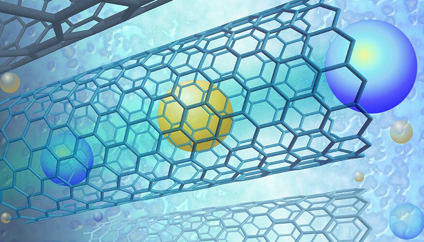
Every year, millions of people around the world die from drinking unclean water. Now, researchers have developed a process that can purify water, no matter how dirty it is, in a single step. Scientists from Australian research organization CSIRO have created a filtration technique using a graphene film with microscopic nano-channels that lets water pass through, but stops pollutants. The process, called “Graphair”, is so effective that water samples from Sydney Harbor were safe to drink after being treated.
And while the film hails from graphene, Graphair is comparatively cheaper, faster and more environmentally-friendly to make, as its primary component is renewable soybean oil, which also helps maximise the efficiency of the purifying technique’s filter counterpart. Over time, oil-based pollutants can impede water filters, so contaminants have to be removed before filtering can even begin, but using Graphair removes these pollutants faster than any other method.
Water purification usually involves a complex process of several steps, so this breakthrough could have a significant impact on the some 2.1 billion people who don’t have clean, safe drinking water. “All that’s needed is heat, our graphene, a membrane filter and a small water pump. We’re hoping to commence field trials in a developing world community next year,” said lead author Dr Dong Han Seo, who added that the team is looking for industry partners to help scale up the technology, and is also working on other applications for Graphair, such as seawater and industrial effluents.
Summary: Nanodocs are medical nanorobots that work from inside like a tiny doctor. The authors of a recent research study say we may be able to swallow the doctor sooner than we think. Once considered science fiction, the ability to “swallow the surgeon” – using medical nanobots to diagnose and treat disease from inside the body – is becoming a reality. The study authors highlight recent advances in nanotechnology tools, such as nanodrillers, microgrippers, and microbullets – and show how nanodocs have tremendous potential in the areas of precision surgery, detection, detoxification and targeted drug delivery. [Cover photo: The old way to swallow the surgeon. Credit: R. Collin Johnson / Attributed to Stanford University.]
Imagine that you need to repair a defective heart valve, a major surgery. Instead of ripping your chest cut open, a doctor merely injects you with a syringe full of medical nanorobots, called nanodocs for short. You emerge from the ‘surgery’ unscathed, and your only external wound is the puncture hole from the injection.
According to a recent study published by nanorobotic engineers at the University of California San Diego (UCSD), the concept of ‘swallow the doctor’ may be closer to reality than we think.
You can’t peer very far down into a well or below the surface of the ocean before things go dark—light does not penetrate to such depths. Though the brain is far from bottomless, neuroscientists face the same lack of light when they try to study living deep-brain structures. This is especially frustrating given that optogenetics, a method for manipulating genetically tagged brain cells with light, has exploded in popularity over the past decade. “Optogenetics has been a revolutionary tool for controlling neurons in the lab, and hopefully someday in the clinic,” says Thomas McHugh, research group leader at the RIKEN Brain Science Institute in Japan. “Unfortunately, delivering light within brain tissue requires invasive optical fibers.”
McHugh and colleagues now have a solution for sending light to new depths in the brain. As they report in Science on February 9, upconversion nanoparticles (UCNPs) can act as a conduit for laser light delivered from outside the skull. These nanoparticles absorb near-infrared laser light and in turn emit visible photons to areas that are inaccessible to standard optogenetics. This method was used to turn on neurons in various brain areas as well as silence seizure activity and evoke memory cells. “Nanoparticles effectively extend the reach of our lasers, enabling the ‘remote’ delivery of light and potentially leading to non-invasive therapies,” says McHugh.
In optogenetics, blue-green light is used to turn neurons on or off via light-responsive ion channels. Light at these wavelengths, however, scatters strongly and is at the other end of the spectrum from the near-infrared light that can penetrate deeper into brain tissue. UCNPs composed of elements from the lanthanide family can act as a bridge. Their ‘optogenetic actuation’ turns low-energy near-infrared laser light into blue or green wavelengths for control of specifically labeled cells. Though such bursts of light deliver considerable energy to a small area, temperature increases or cellular damage were not observed.
Spin a merry-go-round fast enough and the riders fly off in all directions. But the spinning particles in a Rice University lab do just the opposite.
Experiments in the Rice lab of chemical engineer Sibani Lisa Biswal show micron-sized spheres coming together under the influence of a rapidly spinning magnetic field. That’s no surprise because the particles themselves are magnetized.
But how they come together is of interest as the particles first gather into a disorganized aggregated cluster and then into a crystal-like regimen as the magnetic field becomes stronger.
Credit: University of New Mexico For years, scientists have long wrestled with the control and manipulation of light, a long-standing scientific ambition with major implications for the development of technology. With the growth in nanophotonics, scientists are making gains faster than ever exploiting structures with dimensions comparable to the wavelength of light. Scientists at The University of New Mexico studying the field of nanophotonics are developing new perspectives never seen before through their research. In turn, the understanding of these theoretical concepts is enabling physic…