Toggle light / dark theme

Marrying two layers of graphene is an easy route to the blissful formation of nanoscale diamond, but sometimes thicker is better.

While it may only take a bit of heat to turn a treated bilayer of the ultrathin material into a cubic lattice of diamane, a bit of in just the right place can convert few-layer graphene as well.

The otherwise chemically driven process is theoretically possible according to scientists at Rice University, who published their most recent thoughts on making high-quality diamane—the 2-D form of diamond—in the journal Small.

Researchers led by Technische Universität Kaiserslautern (TUK) and the University of Vienna successfully constructed a basic building block of computer circuits using magnons to convey information, in place of electrons. The ‘magnonic half-adder’ described in Nature Electronics, requires just three nanowires, and far less energy than the latest computer chips.

A team of physicists are marking a milestone in the quest for smaller and more energy-efficient computing: they developed an integrated circuit using magnetic material and magnons to transmit binary data, the 1s and 0s that form the foundation of today’s computers and smartphones.

The new circuit is extremely tiny, with a streamlined, 2-D design that requires about 10 times less energy than the most advanced computer chips available today, which use CMOS technology. While the current magnon configuration is not as fast as CMOS, the successful demonstration can now be explored further for other applications, such as quantum or neuromorphic computing.

Imagine a mobile phone charger that doesn’t need a wireless or mains power source. Or a pacemaker with inbuilt organic energy sources within the human body.

Australian researchers led by Flinders University are picking up the challenge of “scavenging” invisible power from low-frequency vibrations in the surrounding environment, including wind, air or even contact-separation energy (static electricity).

“These so-called triboelectric nanogenerators (or TENGs) can be made at low cost in different configurations, making them suitable for driving such as personal electronics (mobile phones), biomechanics devices (pacemakers), sensors (temperature/pressure/chemical sensors), and more,” says Professor Youhong Tang, from Flinders University’s College of Science and Engineering.

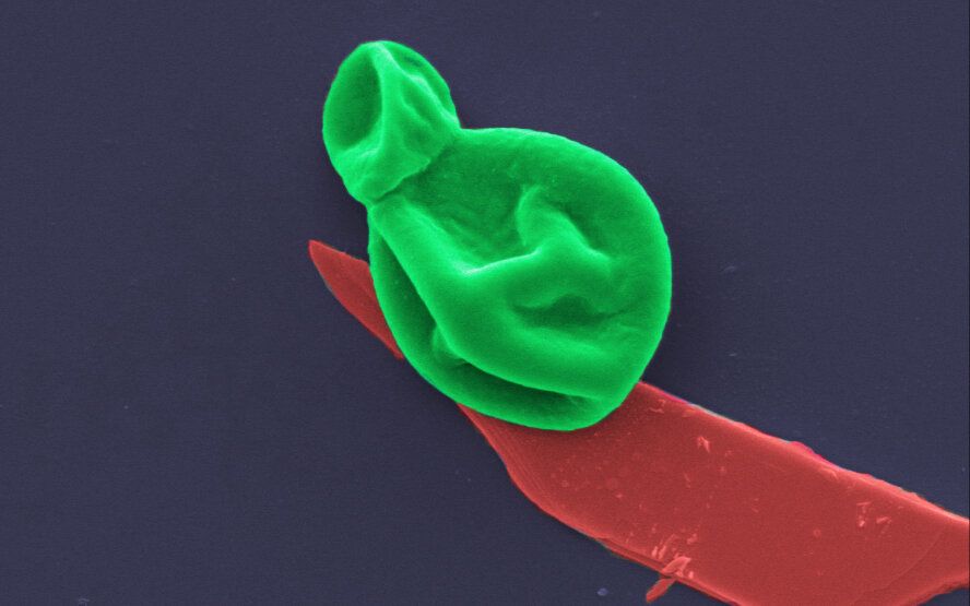
A group of scientists at Northeastern University are making progress using nanotechnology to prevent, diagnose and fight the coronavirus.

Thomas Webster, professor of chemical engineering at Northeastern University, has been working with nanotechnology for decades. Now, he and his team are finding new applications with the coronavirus.

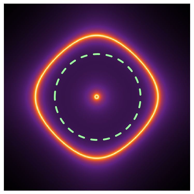
Researchers have developed a method to ‘squeeze’ visible light in order to see inside tiny memory devices. The technique will allow researchers to probe how these devices break down and how their performance can be improved for a range of applications.

The team, led by the University of Cambridge, used the technique to investigate the materials used in random access memories, while in operation. The results, reported in the journal Nature Electronics, will allow detailed study of these materials, which are used in devices.

The ability to understand how structural changes characterize the function of these materials, which are used for , ultra-responsive devices called memristors, is important to improve their performance. However, looking inside the 3D nanoscale devices is difficult using traditional techniques.

Could make awesome computers.


Materials scientists who work with nano-sized components have developed ways of working with their vanishingly small materials. But what if you could get your components to assemble themselves into different structures without actually handling them at all?

Verner Håkonsen works with cubes so tiny that nearly 5 billion of them could fit on a pinhead.

He cooks up the cubes in the NTNU NanoLab, in a weird-looking glass flask with three necks on the top using a mixture of chemicals and special soap.

Virtual reality software which allows researchers to ‘walk’ inside and analyse individual cells could be used to understand fundamental problems in biology and develop new treatments for disease.

The software, called vLUME, was created by scientists at the University of Cambridge and 3D image analysis software company Lume VR Ltd. It allows super-resolution microscopy data to be visualised and analysed in virtual reality, and can be used to study everything from individual proteins to entire cells. Details are published in the journal Nature Methods.

Super-resolution microscopy, which was awarded the Nobel Prize for Chemistry in 2014, makes it possible to obtain images at the nanoscale by using clever tricks of physics to get around the limits imposed by light diffraction. This has allowed researchers to observe molecular processes as they happen. However, a problem has been the lack of ways to visualise and analyse this data in three dimensions.