A recent study published in the Cell Chemical Biology Journal described a small molecule inhibitor of proliferating cell nuclear antigen (PCNA) that selectively kills cancer cells.
Study: Small molecule targeting of transcription-replication conflict for selective chemotherapy. Image Credit: Lightspring/Shutterstock.com.
These and other missions on the horizon will face the same obstacle that has plagued scientists since they first attempted to search for signs of Martian biology with the Viking landers in the 1970s: There is no definitive signature of life.
That might be about to change. In 2021, a team led by Lee Cronin of the University of Glasgow in Scotland and Sara Walker of Arizona State University proposed a very general way to identify molecules made by living systems—even those using unfamiliar chemistries. Their method, they said, simply assumes that alien life forms will produce molecules with a chemical complexity similar to that of life on Earth.
Called assembly theory, the idea underpinning the pair’s strategy has even grander aims. As laid out in a recentseries of publications, it attempts to explain why apparently unlikely things, such as you and me, even exist at all. And it seeks that explanation not, in the usual manner of physics, in timeless physical laws, but in a process that imbues objects with histories and memories of what came before them. It even seeks to answer a question that has perplexed scientists and philosophers for millennia: What is life, anyway?
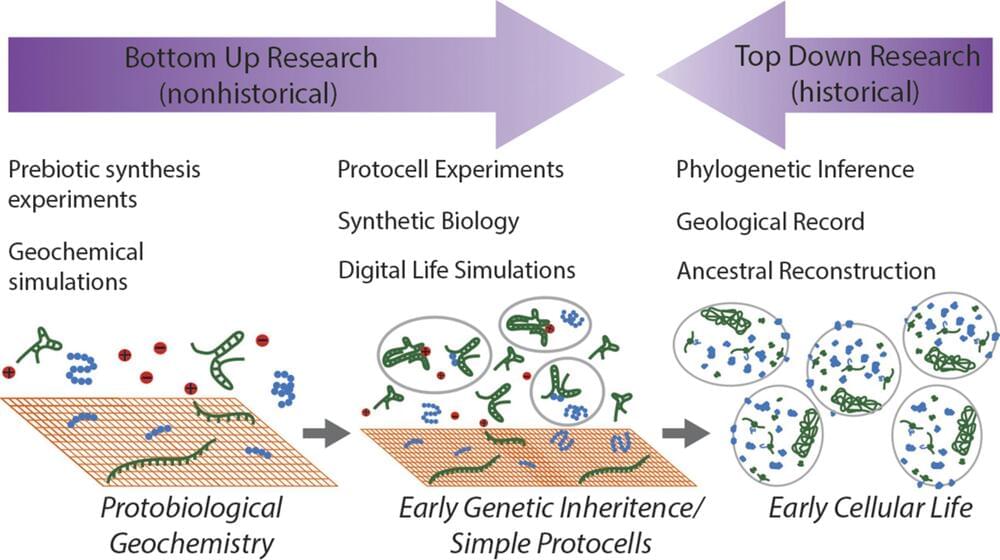
The origin and early evolution of life is generally studied under two different paradigms: bottom up and top down. Prebiotic chemistry and early Earth geochemistry allow researchers to explore possible origin of life scenarios. But for these “bottom–up” approaches, even successful experiments only amount to a proof of principle. On the other hand, “top–down” research on early evolutionary history is able to provide a historical account about ancient organisms, but is unable to investigate stages that occurred during and just after the origin of life. Here, we consider ancient electron transport chains (ETCs) as a potential bridge between early evolutionary history and a protocellular stage that preceded it. Current phylogenetic evidence suggests that ancestors of several extant ETC components were present at least as late as the last universal common ancestor of life. In addition, recent experiments have shown that some aspects of modern ETCs can be replicated by minerals, protocells, or organic cofactors in the absence of biological proteins. Here, we discuss the diversity of ETCs and other forms of chemiosmotic energy conservation, describe current work on the early evolution of membrane bioenergetics, and advocate for several lines of research to enhance this understanding by pairing top–down and bottom–up approaches.
In this “Ask Me Anything” (AMA) episode, Peter delves into the realm of genetics, unraveling its connection to disease and emphasizing the value of understanding one’s genetic risks. He elucidates essential background knowledge on genetics before delving into the myriad reasons why individuals might consider genetic testing. Peter differentiates scenarios where genetic testing provides genuine insights from those where it may not be as useful. From there, Peter explores a comprehensive comparison of commercial direct-to-consumer genetic tests, providing insights on interpreting results and identifying the standout options for gaining insights into personal health.
In this sneak peek, we discuss: 00:00 — Intro. 02:09 — Defining the term “genetics” and why it’s important. 04:03 — What is DNA, and how does it impact our biology and traits? 07:13 — How are genetics passed down from parent to child? 11:44 — How much do genes vary across individuals? 16:22 — Which traits are determined by genetics versus experience or environmental factors? 22:30 — Reasons for genetic testing.
In the full episode, we also discuss: –What exactly is being measured by a genetic test?; –Testing for monogenic disorders; –Understanding polygenic risk; –Is genetic testing more important for someone who doesn’t know their family history?; –What does it mean to be positive for a particular variant?; –What does it mean to be negative for a particular variant?; –How does someone get genetic testing through their healthcare provider, and how are these tests performed?; –The financial cost of various genetic tests; –Could having a risk allele for a disease result in an increase in one’s insurance premium?; –Other risks associated with genetic testing; –How do commercial, direct-to-consumer genetic tests compare to the information one might receive from clinical genetic testing?; –Are certain direct-to-consumer tests better than others?; –How long until whole genome sequencing becomes genuinely useful?; –How useful are personalized dietary recommendations based on genetics?; –Final thoughts and advice regarding genetic testing; and. –More.
——- About:
The Peter Attia Drive is a deep-dive podcast focusing on maximizing longevity, and all that goes into that from physical to cognitive to emotional health. With over 70 million episodes downloaded, it features topics including exercise, nutritional biochemistry, cardiovascular disease, Alzheimer’s disease, cancer, mental health, and much more.
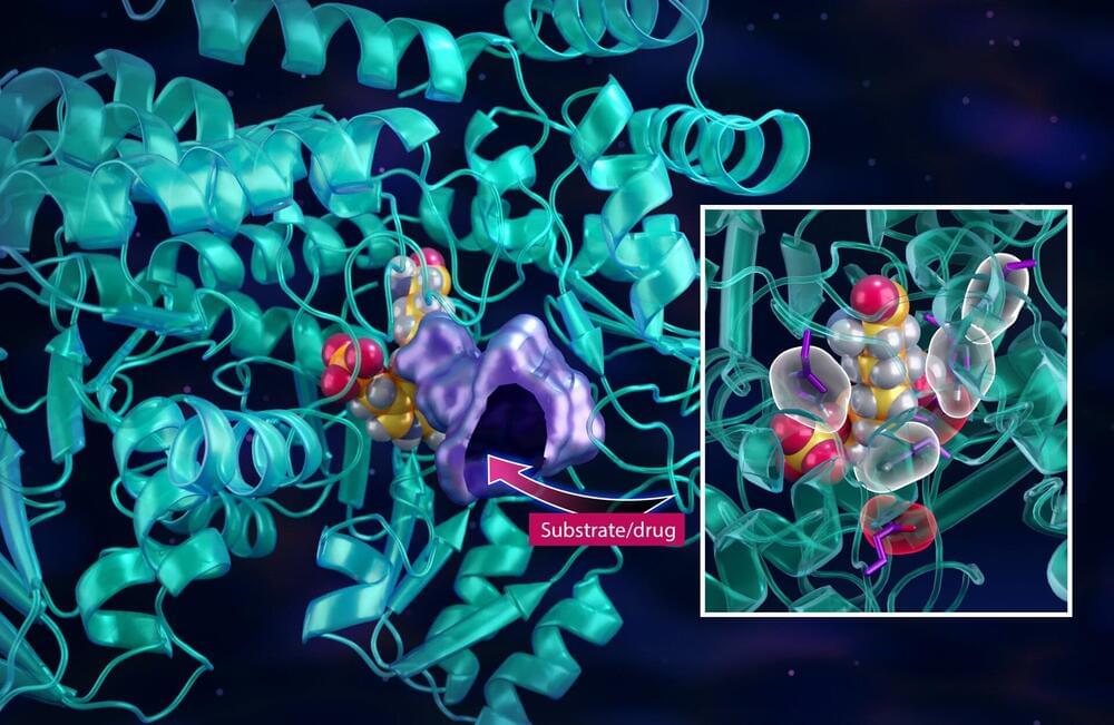
After a highly lauded research campaign that successfully redesigned a hepatitis C drug into one of the leading drug treatments for COVID-19, scientists at the Department of Energy’s Oak Ridge National Laboratory are now turning their drug design approach toward cancer.
In their latest study, published in the journal Communications Chemistry, the team used neutrons and X-rays to draw a roadmap of every atom, chemical bond and electrical charge inside a key enzyme that belongs to a metabolic pathway that cancer cells dramatically overuse to reproduce.
This new information essentially helps pave the way for developing new drugs that act as roadblocks along the metabolic pathway to cut off the supply of vital resources to cancers cells. The drugs would be designed to target highly aggressive tumor-forming cancers that too often become terminal such as lung, colon, breast, pancreatic and prostate cancers.
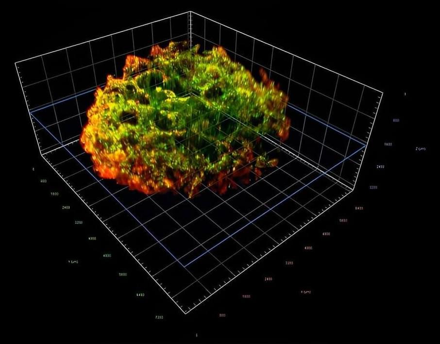
Tumor cells are known to be fickle sleeper agents, often lying dormant in distant tissues for years before reactivating and forming metastasis. Numerous factors have been studied to understand why the activation occurs, from cells and molecules to other components in the so-called tissue microenvironment.
Now, an interdisciplinary Cornell University team has identified a new mechanism regulating tumor growth in the skeleton, the primary site of breast cancer metastasis: mineralization of the bone matrix, a fibrous mesh of organic and inorganic components that determines the unique biochemical and biomechanical properties of our skeleton.
The team’s paper, “Bone-Matrix Mineralization Dampens Integrin-Mediated Mechanosignalling and Metastatic Progression in Breast Cancer,” published Aug. 7 in Nature Biomedical Engineering. The co-lead authors are research associate Siyoung Choi and doctoral student Matthew Whitman.
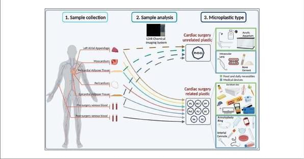
Microplastics have been detected in human stool, lungs, and placentas, which have direct exposure to the external environment through various body cavities, including the oral/anal cavity and uterine/vaginal cavity. Crucial data on microplastic exposure in completely enclosed human organs are still lacking. Herein, we used a laser direct infrared chemical imaging system and scanning electron microscopy to investigate whether microplastics exist in the human heart and its surrounding tissues. Microplastic specimens were collected from 15 cardiac surgery patients, including 6 pericardia, 6 epicardial adipose tissues, 11 pericardial adipose tissues, 3 myocardia, 5 left atrial appendages, and 7 pairs of pre-and postoperative venous blood samples.
Alien enthusiasts have a new reason to get excited about potential life on Mars, after scientists found cracked mud on the Red Planet.
A recent research paper showed that the conditions that created cracks in the surface of Mars might have been favourable for microscopic life to thrive.
While scientists don’t yet know how life on Earth began, a prevalent theory is that repeated cycles of wet and dry conditions might have helped build the complex chemical building blocks needed for microbial life.
An international team that includes researchers from the University of Toronto has designed and implemented a new model for photoreactors, a solar-powered technology for converting water, carbon dioxide, methane and nitrogen into greener chemicals and fuels.
The innovative design allows the photoreactor to capture photons at high efficiency under varying sun directions, eliminating the need for sun-tracking. The panels are also manufacturable via extrusion of polymers, making them inexpensive and easily manufacturable at scale—all of which could help make a sustainable future more affordable and practical.
Geoffrey Ozin, University Professor in U of T’s department of chemistry in the Faculty of Arts & Science, and his team collaborated with researchers from the Karlsruhe Institute of Technology (KIT) in Germany on the project.


