Toggle light / dark theme

How Depression Affects The Brain — Yale Medicine Explains

For more information on mental health or #YaleMedicine, visit: https://www.yalemedicine.org/conditions/topics/mental-health.

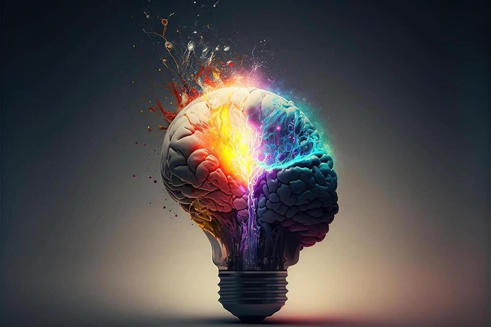
For many people, depression turns out to be one of the most disabling illnesses that we have in society. Despite the treatments that we have available, many people are not responding that well. It’s a disorder that can be very disabling in society. It’s also a disorder that has medical consequences. By understand the neurobiology of depression we hope to be able more to find the right treatment for the patient suffering from this disease. The current standard of care for the treatment of depression is based on what we call the monoamine deficiency hypothesis. Essentially, presuming that one of three neurotransmitters in the brain is deficient or underactive. But the reality is, there are more than 100 neurotransmitters in the brain. And billions of connections between neurons. So we know that that’s a limited hypothesis. Neurotransmitters can be thought of as the chemical messengers within the brain, it’s what helps one cell in the brain communicate with another, to pass that message along from one brain region to another. For decades, we thought that the primary pathology, the primary cause of depression was some abnormality in these neurotransmitters, specifically serotonin or norepinephrine. However, norepinephrine and serotonin did not seem to be able to account for this cause, or to cause the symptoms of depression in people who had major depression. Instead, the chemical messengers between the nerve cells in the higher centers of the brain, which include glutamate and GABA, were possibilities as alternative causes for the symptoms of depression. When you’re exposed to severe and chronic stress like people experience when they have depression, you lose some of the connections between the nerve cells. The communication in these circuits becomes inefficient and noisy, we think that the loss of these synaptic connections contributes to the biology of depression. There are clear differences between a healthy brain and a depressed brain. And the exciting thing is, when you treat that depression effectively, the brain goes back to looking like a healthy brain, both at the cellular level and at a global scale. It’s critical to understand the neurobiology of depression and how the brain plays a role in that for two main reasons. One, it helps us understand how the disease develops and progresses, and we can start to target treatments based on that. We are in a new era of psychiatry. This is a paradigm shift, away from a model of monoaminergic deficiency to a fuller understanding of the brain as a complex neurochemical organ. All of the research is driven by the imperative to alleviate human suffering. Depression is one of the most substantial contributors to human suffering. The opportunity to make even a tiny dent in that is an incredible opportunity.

Tiny Robots Detect and Treat Cancer by Traveling Deep into the Lungs

A tiny robot which can travel deep into the lungs to detect and treat the first signs of cancer has been developed by researchers at the University of Leeds. The ultra-soft tentacle, which measures just two millimeters in diameter, and is controlled by magnets, can reach some of the smallest bronchial tubes and could transform the treatment of lung cancer. The researchers tested the magnetic tentacle robot on the lungs of a cadaver and found that it can travel 37 percent deeper than the standard equipment and leads to less tissue damage. It paves the way for a more accurate, tailored, and far less invasive approach to treatment.

The work is published in Nature Engineering Communications in the paper, “Magnetic personalized tentacles for targeted photothermal cancer therapy in peripheral lungs.

“This new approach has the advantage of being specific to the anatomy, softer than the anatomy and fully-shape controllable via magnetics,” notes Pietro Valdastri, PhD, director of the Science and Technologies Of Robotics in Medicine (STORM) Lab at the University of Leeds. “These three main features have the potential to revolutionize navigation inside the body.”

Stanford Medicine researchers take part in HuBMAP, showing what healthy human tissue looks like

More often than not, studies of human biology are conducted when the body is under duress from infection or disease. Now, as part of a larger effort to delineate what “healthy” looks like, two Stanford Medicine teams have unfurled detailed molecular maps of healthy human intestinal and placental tissues. The maps, which capture cell types, cell quantity and other cellular nuances, are just two of a collection of maps that will establish a cellular baseline for the majority of the human body, including where cells in certain tissues congregate, how tissues develop during pregnancy and how cell-to-cell interactions drive human biology.

The studies, which published in Nature on July 19, are part of a larger effort spearheaded by the Human Biomolecular Atlas Program — called HuBMAP — funded by the National Institutes of Health. It aims to fill gaps in our knowledge of how the human body works when it’s in tip-top shape. Dozens of teams from the United States and Europe contribute to the HuBMAP consortium.

“In research, we have a habit of studying things that are abnormal without really understanding what normal looks like,” said Michael Angelo, MD, PhD, an assistant professor of pathology who is also the co-chair of the HuBMAP steering committee. “That’s created a big gap in our knowledge. HuBMAP is the only effort that is systematically focusing on the spatial architecture of these tissues.”

Simple Brain Hack Could Boost Learning and Improve Mental Health

Adopting a curious mindset over a high-pressure one can enhance memory, according to recent research from Duke University. The study showed that participants who envisioned themselves as a thief planning a heist in a virtual art museum demonstrated better recall of the paintings they encountered than those who imagined executing the heist on the spot while playing the same computer game.

The slight variation in motivations — the urgent need to achieve immediate goals versus the curious exploration for future objectives — could have significant implications in real-life scenarios. These include incentivizing people to receive a vaccine, prompting action against climate change, and potentially providing new treatments for psychiatric conditions.

The findings were recently published in the Proceedings of the National Academy of Sciences.

Boning Up on the Unique Genetics of the Human Skeletal System

Humans have a distinctive skeleton, and are the only bipedal great apes (the great ape species are bonobos, chimpanzees, gorillas, orangutans, and humans). While the evolution of the human skeleton enabled us to walk upright, it also led to the rise of musculoskeletal disease. It’s thought that cognitive development began to accelerate in humans once we started to move around, adapt to new environments, and make use of tools. Researchers have now used advanced computational tools and a trove of human genetic data in the UK Biobank to outline the genetic changes that occurred as primates started to walk upright for the first time.

These findings, which were reported in Science, have suggested that natural selection had a strong influence on the genetic changes that altered our anatomy, and gave early humans an evolutionary leg up.

Study explains link to increased cardiovascular risks for people with obstructive sleep apnea

Researchers have found that people with obstructive sleep apnea have an increased cardiovascular risk due to reduced blood oxygen levels, largely explained by interrupted breathing. Obstructive sleep apnea has long been associated with increased risk of cardiovascular issues, including heart attack, stroke, and death, but the findings from this study, published in the American Journal of Respiratory and Critical Care Medicine, show the mechanism mostly responsible for the link.

“These findings will help better characterize high-risk versions of obstructive ,” said Ali Azarbarzin, Ph.D., a study author and director of the Sleep Apnea Health Outcomes Research Group at Brigham and Women’s Hospital and Harvard Medical School, Boston. “We think that including a higher-risk version of obstructive sleep apnea in a would hopefully show that treating sleep apnea could help prevent future cardiovascular outcomes.”

Researchers reviewed data from more than 4,500 middle-aged and older adults who participated in the Osteoporotic Fractures in Men Study (MrOS) and the Multi-Ethnic Study of Atherosclerosis (MESA), and sought to identify features of obstructive sleep apnea that could explain why some people were more likely than others to develop cardiovascular disease or related death.

/* */