AI can spot patterns in the data from blood tests that can give an early warning of disease.
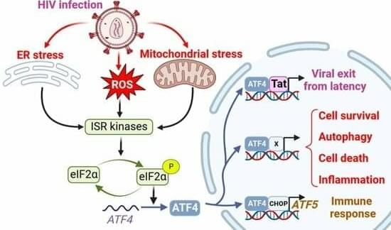
Cellular integrated stress response (ISR), the mitochondrial unfolded protein response (UPRmt), and IFN signaling are associated with viral infections. Activating transcription factor 4 (ATF4) plays a pivotal role in these pathways and controls the expression of many genes involved in redox processes, amino acid metabolism, protein misfolding, autophagy, and apoptosis. The precise role of ATF4 during viral infection is unclear and depends on cell hosts, viral agents, and models. Furthermore, ATF4 signaling can be hijacked by pathogens to favor viral infection and replication. In this review, we summarize the ATF4-mediated signaling pathways in response to viral infections, focusing on human immunodeficiency virus 1 (HIV-1). We examine the consequences of ATF4 activation for HIV-1 replication and reactivation.
Understanding the behavior of the molecules and cells that make up our bodies is critical for the advancement of medicine. This has led to a continual push for clear images of what is happening beyond what the eye can see. In a study recently published in Science Advances, researchers from Osaka University have reported a method that gives high-resolution Raman microscopy images.
Raman microscopy is a useful technique for imaging biological samples because it can provide chemical information about specific molecules—such as proteins—that take part in the body’s processes. However, the Raman light that comes from biological samples is very weak, so the signal can often get swamped by the background noise, leading to poor images.
The researchers have developed a microscope that can maintain the temperature of previously frozen samples during the acquisition. This has allowed them to produce images that are up to eight times brighter than those previously achieved with Raman microscopy.
Link :
Imagine a life where your body’s internal “battery” runs low every single day, demanding constant recharging just to keep going. For millions of people living with Type 1 diabetes, this is the exhausting reality—one where insulin injections act as the lifeline, replacing what the body can no longer produce on its own. But what if the body could be taught to recharge itself again?
In a world-first medical breakthrough, this question has moved from possibility to reality. A woman’s own stem cells have been successfully used to reverse her Type 1 diabetes, a condition once thought to be irreversible. Scientists turned her blood stem cells into insulin-producing powerhouses, effectively “rebooting” her pancreas and allowing her body to produce insulin naturally for the first time in years.
This achievement isn’t just a milestone for one patient; it’s a bold step toward a future where Type 1 diabetes may no longer require a lifetime of management. So, how did this groundbreaking transformation happen? And what does it mean for millions waiting for a cure?


Monitoring electrical signals in biological systems helps scientists understand how cells communicate, which can aid in the diagnosis and treatment of conditions like arrhythmia and Alzheimer’s.
But devices that record electrical signals in cell cultures and other liquid environments often use wires to connect each electrode on the device to its respective amplifier. Because only so many wires can be connected to the device, this restricts the number of recording sites, limiting the information that can be collected from cells.
MIT researchers have now developed a biosensing technique that eliminates the need for wires. Instead, tiny, wireless antennas use light to detect minute electrical signals.
Leveraging the success of this new program, just about two years from its launch DeepMind’s AI spinout Isomorphic announced two drug discovery deals, worth $3 billion each, with Eli Lilly and Novartis.
Earlier this year, microprocessor giant NVIDIA also dove head first into AI for drug discovery, making big investments and deals with leaders like Recursion Pharmaceuticals and Genentech.
AI in drug discovery seems to be having a moment.

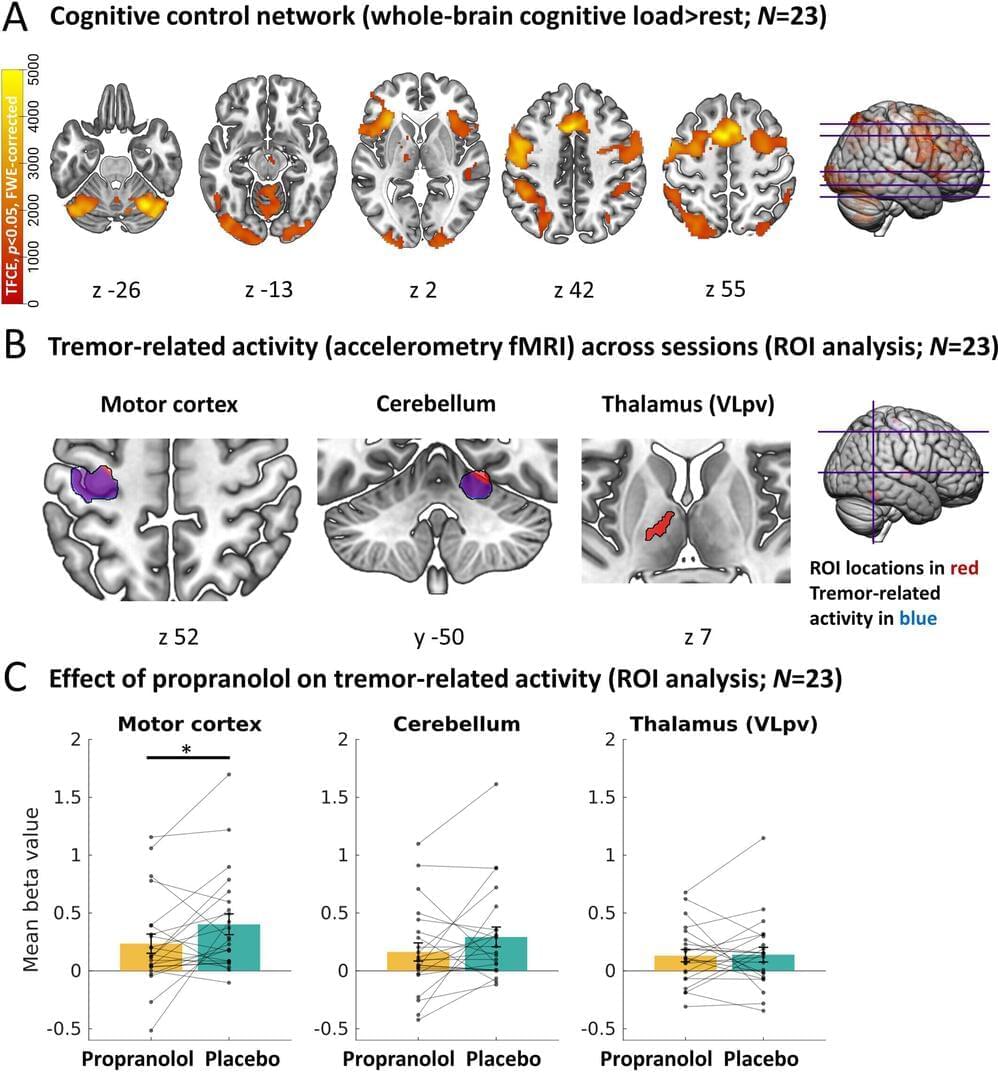
The standard medication levodopa does not always work against tremors in Parkinson’s disease, especially in stressful situations. Propranolol, however, does work during stress, providing insight into the role of the stress system in tremors. MRI scans reveal that propranolol directly inhibits activity in the brain circuit that controls tremors. Doctors may consider this medication when levodopa is ineffective.
People with Parkinson’s disease report that tremors worsen during stressful situations. “Tremors act as a sort of barometer for stress; you see this in all people with Parkinson’s,” says neurologist Rick Helmich from Radboud university medical center.
The commonly used drug levodopa usually helps with tremors, but it tends to be less effective during stress, when tremors are often at their worst. Helmich and his team wanted to investigate whether a medication targeting the stress system could help and how this effect of stress on tremors works in the brain. The work is published in the journal Annals of Neurology.
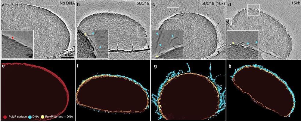
In a discovery that could redefine how we understand cellular resilience and adaptability, scientists at Scripps Research have unlocked the secret interactions between a primordial inorganic polymer of phosphate known as polyphosphate (polyP), and two basic building blocks of life: DNA and the element magnesium. These components formed clusters of tiny liquid droplets–also known as condensates–with flexible and adaptable structures.
PolyP and magnesium are involved in many biological processes. Thus, the findings could lead to new methods for tuning cellular responses, which could have impactful applications in translational medicine.
The ensuing study, published in Nature Communications on October 26, 2024, reveals a delicate “Goldilocks” zone—a specific magnesium concentration range—where DNA wraps around polyP-magnesium ion condensates. Similar to a thin eggshell covering a liquid-like interior, this seemingly simple structure may help cells organize and protect their genetic material.