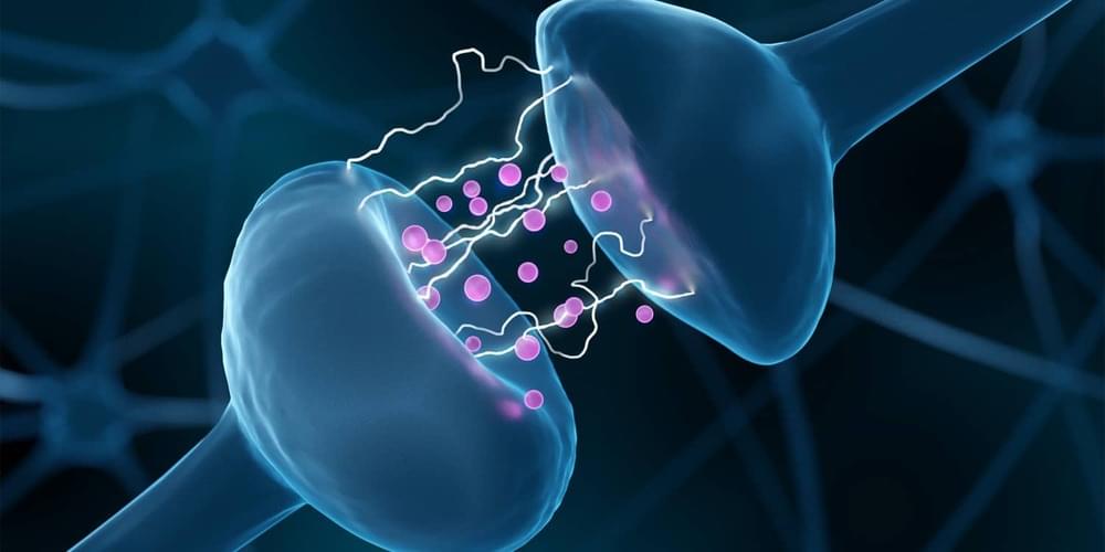
Toggle light / dark theme

Computational modelling of the brain requires accurate representation of the tissues concerned. Mechanical testing has numerous challenges, in particular for low strain rates, like neurosurgery, where redistribution of fluid is biomechanically important. A finite-element (FE) model was generated in FEBio, incorporating a spring element/fluid–structure interaction representation of the pia–arachnoid complex (PAC). The model was loaded to represent gravity in prone and supine positions. Material parameter identification and sensitivity analysis were performed using statistical software, comparing the FE results to human in vivo measurements. Results for the brain Ogden parameters µ, α and k yielded values of 670 Pa, −19 and 148 kPa, supporting values reported in the literature. Values of the order of 1.2 MPa and 7.7 kPa were obtained for stiffness of the pia mater and out-of-plane tensile stiffness of the PAC, respectively. Positional brain shift was found to be non-rigid and largely driven by redistribution of fluid within the tissue. To the best of our knowledge, this is the first study using in vivo human data and gravitational loading in order to estimate the material properties of intracranial tissues. This model could now be applied to reduce the impact of positional brain shift in stereotactic neurosurgery.

Finite-element (FE)-based computational models of the human brain are an increasingly common research tool, with applications ranging from head impact to neurosurgery. Studies considering head impacts are generally concerned with traumatic brain injury (TBI), where a better understanding of the underlying mechanisms is essential for the development of prevention measures [1]. Within neurosurgery, efforts are primarily focused on tumour resection, where loss of cerebrospinal fluid (CSF) and tissue resection are responsible for much of the deformation [2]. Movement and deformation of the intact brain, known as brain shift, is clinically significant in stereotactic neurosurgical procedures such as deep brain stimulation where electrode placement accuracy correlates with patient outcomes [3].

Aging remains a primary risk factor for a host of diseases, including leading causes of death. Aging and associated diseases are inherently multifactorial, with numerous contributing factors and phenotypes at the molecular, cellular, tissue, and organismal scales. Despite the complexity of aging phenomena, models currently used in aging research possess limitations. Frequently used in vivo models often have important physiological differences, age at different rates, or are genetically engineered to match late disease phenotypes rather than early causes. Conversely, routinely used in vitro models lack the complex tissue-scale and systemic cues that are disrupted in aging.

San Diego based biotech Immunicom has developed an innovative therapeutic device that is intended to remove specific factors from blood that prevent our immune systems from fighting disease. Existing “therapeutic apheresis” treatments usually involve modification to cells harvested from patients or some form of plasma exchange. Immunicom’s technology uses a similar treatment procedure but says its approach of “capturing” targeted molecules is very different from other treatments.

Immunicom creates customised molecules that are designed to capture specific proteins or cytokines, but instead of turning those into drugs, it deploys them via its proprietary cartridges, which are essentially small liquid filters that can be fitted into any plasmapheresis machine. In a treatment similar to dialysis, the cartridges are used to treat a patient’s blood and plasma outside their body, reducing the levels of the target factors, with the goal of enabling the immune system to fight disease.

While Immunicom has focused its initial efforts on treatments for cancer, the company claims its technology can also potentially be applied to longevity and the treatment of a wide range of age-related diseases.

A study published Wednesday in the JAMA Psychiatry journal shows that four key genetic variations are more common in military veterans who have taken their own life or considered it.

Scientists from Duke University in Durham, North Carolina, found the pattern while analyzing blood samples from a database that included 633,778 U.S. veterans, cross-referenced with the International Suicide Genetics Consortium of more than 549,000 individuals.

The obtained samples were sequenced to create genetic profiles compared to participants’ medical records, showing that 121,211 recorded cases of attempted suicide or thoughts about killing themselves.