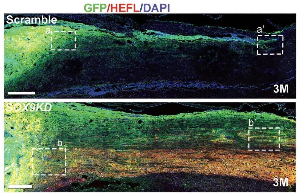
Toggle light / dark theme

The ‘unknome’: A database of human genes we know almost nothing about

Researchers from the United Kingdom hope that a new, publicly available database they have created will shrink, not grow, over time. That’s because it is a compendium of the thousands of understudied proteins encoded by genes in the human genome, whose existence is known but whose functions are mostly not.

The , dubbed the “unknome,” is the work of Matthew Freeman of the Dunn School of Pathology, University of Oxford, England, and Sean Munro of MRC Laboratory of Molecular Biology in Cambridge, England, and colleagues, and is described in the open access journal PLOS Biology. Their own investigations of a subset of proteins in the database reveal that a majority contribute to important cellular functions, including development and resilience to stress.

The sequencing of the has made it clear that it encodes thousands of likely sequences whose identities and functions are still unknown. There are multiple reasons for this, including the tendency to focus scarce research dollars on already-known targets, and the lack of tools, including antibodies, to interrogate cells about the function of these proteins. But the risks of ignoring these proteins are significant, the authors argue, since it is likely that some, perhaps many, play important roles in critical cell processes, and may both provide insight and targets for therapeutic intervention.

Researchers use quantum circuit to identify single nucleotides

DNA sequencing technology, i.e., determining the order of nucleotide bases in a DNA molecule, is central to personalized medicine and disease diagnostics, yet even the fastest technologies require hours, or days, to read a complete sequence. Now, a multi-institutional research team led by The Institute of Scientific and Industrial Research (SANKEN) at Osaka University, has developed a technique that could lead to a new paradigm for genomic analysis.

DNA sequences are sequential arrangements of the nucleotide bases, i.e., the four letters that encode information invaluable to the proper functioning of an organism. For example, changing the identity of just one nucleotide out of the several billion nucleotide pairs in the can lead to a serious medical condition. The ability to read DNA sequences quickly and reliably is thus essential to some urgent point-of-care decisions, such as how to proceed with a particular chemotherapy treatment.

Unfortunately, genome analysis remains challenging for , and it’s in this context that quantum computers show promise. Quantum computers use quantum bits instead of the zeroes and ones of classical computers, facilitating an exponential increase in computational speed.

Tracing maternal behavior to brain immune function

Immune system changes in the pregnant body that protect the fetus appear to extend to the brain, where a decrease in immune cells late in gestation may factor into the onset of maternal behavior, new research in rats suggests.

In adult female rats that had never given birth—which typically don’t like being around babies—depletion of these cells sped up their care for rat newborns that were placed in their cage.

The loss of these cells, called microglia, and the related uptick in motherly attentiveness were also associated with changes to in several regions of the rat brain, suggesting shifts in have a role in regulating .

A potential strategy to control MAPK4-dependent cancer growth

However, there are no drugs that specifically block MAPK4 that could be tested to reduce tumor growth. Instead, Yang and his colleagues explored an alternative approach.

We showed that blocking both AKT and PDK1 effectively repressed MAPK4-induced cancer cell growth, suggesting a potential therapeutic strategy to treat MAPK4-dependent cancers, such as a subset of TNBC, prostate and lung cancer.

“In this study we have not only advanced our understanding of the molecular mechanism underlying the tumor-promoting activity of MAPK4, we also have found a potential novel therapeutic approach for human cancers,” Yang said.

How Neurons Make Connections

For many people, they are tiny pests. These fruit flies that sometimes hover over a bowl of peaches or a bunch of bananas. But for a dedicated community of researchers, fruit flies are an excellent model organism and source of information into how neurons self-organize during the insect’s early development and form a complex, fully functioning nervous system.

That’s the scientific story on display in this beautiful image of a larval fruit fly’s developing nervous system. Its subtext is: fundamental discoveries in the fruit fly, known in textbooks as Drosophila melanogaster, provide basic clues into the development and repair of the human nervous system. That’s because humans and fruit flies, though very distantly related through the millennia, still share many genes involved in their growth and development. In fact, 60 percent of the Drosophila genome is identical to ours.

Once hatched, as shown in this image, a larval fly uses neurons (magenta) to sense its environment. These include neurons that sense the way its body presses against the surrounding terrain, as needed to coordinate the movements of its segmented body parts and crawl in all directions.

Ancient DNA reveals an early African origin of cattle in the Americas

Cattle may seem like uniquely American animals, steeped in the lore of cowboys, cattle drives and sprawling ranches. But cattle didn’t exist on the American continents prior to the arrival of the Spanish, who brought livestock with them from Europe by way of the Canary Islands.

In a new study, researchers analyzed ancient DNA from Spanish settlements in the Caribbean and Mexico. Their results indicate were also imported from Africa early in the process of colonization, more than 100 years before their arrival was officially documented.

Records kept by Portuguese and Spanish colonists reference breeds from the Andalusian region of Spain but make no mention of transporting cattle from Africa. Some historians have interpreted this omission to mean that the first wave of colonists relied entirely on a small stock of European cattle initially shipped to the Caribbean Islands.

Discovery in nanomachines within living organisms — cytochromes P450 (CYP450s) unleashed as living soft robots

Study reveals an important discovery in the realm of nanomachines within living systems. Prof. Sason Shaik from the Hebrew University of Jerusalem and Dr. Kshatresh Dutta Dubey from Shiv Nadar University, conducted molecular-dynamics simulations of Cytochromes P450 (CYP450s) enzymes, revealing that these enzymes exhibit unique soft-robotic properties.

Cytochromes P450 (CYP450s) are enzymes found in living organisms and play a crucial role in various biological processes, particularly in the metabolism of drugs and xenobiotics. The researchers’ simulations demonstrated that CYP450s possess a fourth dimension — the ability to sense and respond to stimuli, making them soft-robot nanomachines in “living matters.”

In the catalytic cycle of these enzymes, a molecule called a substrate binds to the enzyme. This leads to a process called oxidation. The enzyme’s structure has a confined space that allows it to act like as a sensor and a soft robot. It interacts with the substrate using weak interactions, like soft impacts. These interactions transfer energy, causing parts of the enzyme and the molecules inside it to move. This movement generates ultimately a special substance called oxoiron species, which serves the enzyme to oxidize a variety of different substances.

Lab-grown RPE cells promise to cure age-related blindness

Retinal pigment epithelial (RPE) cells grown on 3D nano scaffolds have the potential to treat age-related macular degeneration, a disease that is making millions of humans blind as they age.

Age-related macular degeneration (AMD) is one of the most common causes of poor eyesight, blurred vision, and blindness in middle and old-age individuals. A team of scientists at Anglia Ruskin University (ARU) has figured out a way to treat this condition using cultured retinal pigment epithelial (RPE) cells.

In their latest study, the ARU team demonstrated a method that allowed them to grow RPE cells on 3D nano scaffolds made of thin nanofibers that can be arranged in any orientation and replicate nerve fibers’ arrangement.