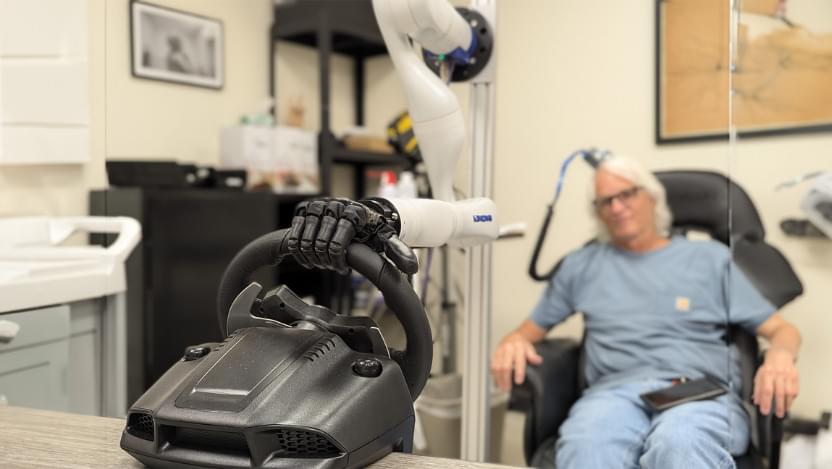
Two new papers document progress in neuroprosthetic technology that lets people feel the shape and movement of objects moving over the “skin” of a bionic hand.
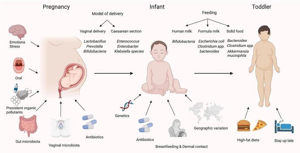
Mechanisms underlying gut microbiota’s role in obesity
Energy absorption and short-chain fatty acids
Gut microbiota regulate energy metabolism through short-chain fatty acids (SCFAs) like acetate, butyrate, and propionate, which are products of fiber fermentation. While butyrate promotes insulin sensitivity and reduces inflammation, propionate may trigger overeating. Dysregulated SCFA production can contribute to obesity by enhancing energy absorption, disrupting appetite regulation, and promoting fat accumulation. Recent findings suggest that modulating SCFA production through dietary interventions can help regulate energy balance and improve metabolic health. Maintaining SCFA balance through diet or microbial modulation holds promise for obesity management.
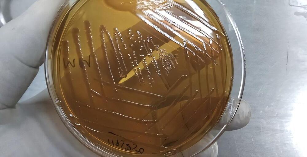
It’s become increasingly clear that the gut microbiome can affect human health, including mental health. Which bacterial species influence the development of disease and how they do so, however, is only just starting to be unraveled.
For instance, some studies have found compelling links between one species of gut bacteria, Morganella morganii, and major depressive disorder. But until now, no one could tell whether this bacterium somehow helps drive the disorder, the disorder alters the microbiome, or something else is at play.
Harvard Medical School researchers have now pinpointed a biologic mechanism that strengthens the evidence that M. morganii influences brain health and provides a plausible explanation for how it does so.
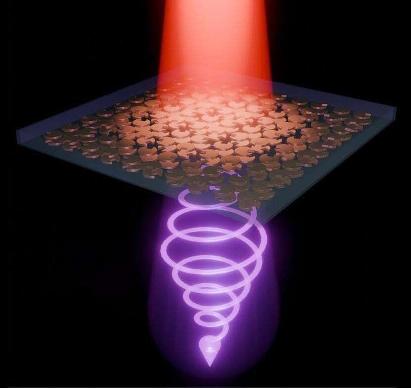
Researchers have developed a revolutionary ultra-thin metasurface that can generate circularly polarized light with remarkable efficiency.
By leveraging the unique properties of chirality and rotational symmetry, this breakthrough eliminates the need for bulky optical setups, enabling more compact and efficient optical devices. This innovation has far-reaching implications for fields such as medical imaging, communications, and quantum physics.
Advancing Optical Technology with Metasurfaces.
Trump—flanked by larry ellison, sam altman, & masayoshi son—announces project stargate.
Trump announces Project Stargate, a $500 billion initiative backed by major tech leaders, aimed at revolutionizing U.S. AI infrastructure, creating jobs, and enhancing healthcare through advanced technologies. AI Infrastructure and Economic Impact.
🏗️Project Stargate, a $500+ billion AI infrastructure initiative, aims to construct colossal data centers and physical campuses across the US, potentially creating over 100,000 American jobs.
🌐The project will build physical and virtual infrastructure to power next-generation AI advancements, with Oracle, SoftBank, and Microsoft as key partners, establishing a new US-centered industry. ## Healthcare Applications.
S AI will enhance healthcare by providing doctors with condition-specific treatment plans based on data from top hospitals like Memorial Sloan Kettering and Stanford. ” + s cancer research focuses on early detection via blood tests, personalized vaccines designed using AI in 48 hours, and robotically-produced mRNA vaccines. ” +## Technological Advancements.
S AI-driven approach promises to accelerate cancer treatment development, potentially leading to unprecedented cure rates. ” + 📊The initiative will leverage AI to improve electronic health records, benefiting patients in underserved areas like Indian River Reservation.
Join us on Patreon! https://www.patreon.com/MichaelLustgartenPhD
Discount Links/Affiliates:
Blood testing (where I get the majority of my labs): https://www.ultalabtests.com/partners/michaellustgarten.
At-Home Metabolomics: https://www.iollo.com?ref=michael-lustgarten.
Use Code: CONQUERAGING At Checkout.
Clearly Filtered Water Filter: https://get.aspr.app/SHoPY
Epigenetic, Telomere Testing: https://trudiagnostic.com/?irclickid=U-s3Ii2r7xyIU-LSYLyQdQ6…M0&irgwc=1
Use Code: CONQUERAGING
NAD+ Quantification: https://www.jinfiniti.com/intracellular-nad-test/
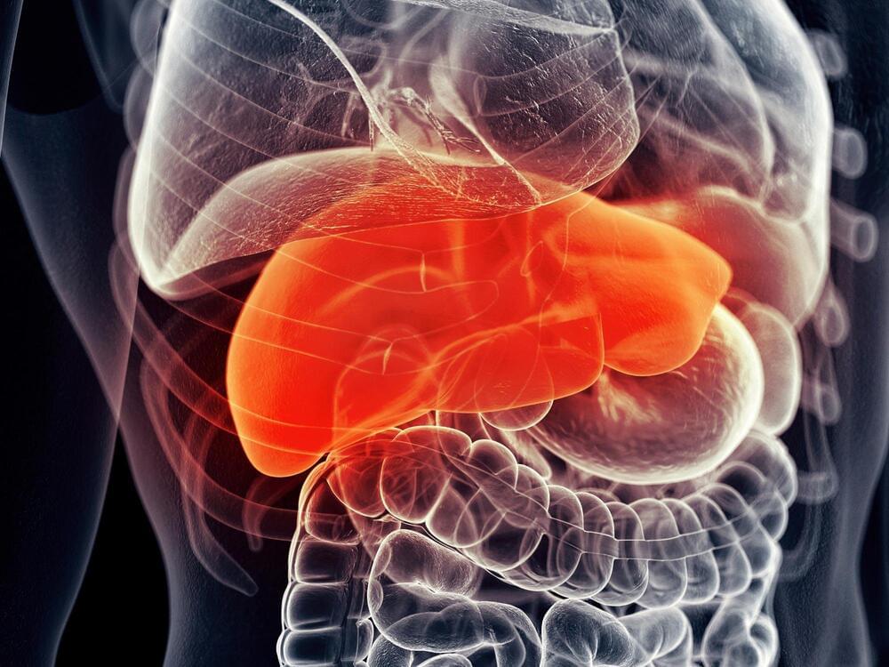
“ tabindex=”0” acid UDCA can regulate tumor growth in mice with liver cancer. This discovery suggests that UDCA dietary supplements could offer a fast and effective way to improve outcomes for liver cancer patients.
Immunotherapy is an advanced cancer treatment that harnesses a patient’s immune system to target and destroy tumors. It has significantly improved outcomes for various cancers, including those of the lung, kidney, and bladder. However, its effectiveness against liver cancer has been notably limited—a concerning issue given that liver cancer rates have nearly tripled over the past 40 years.