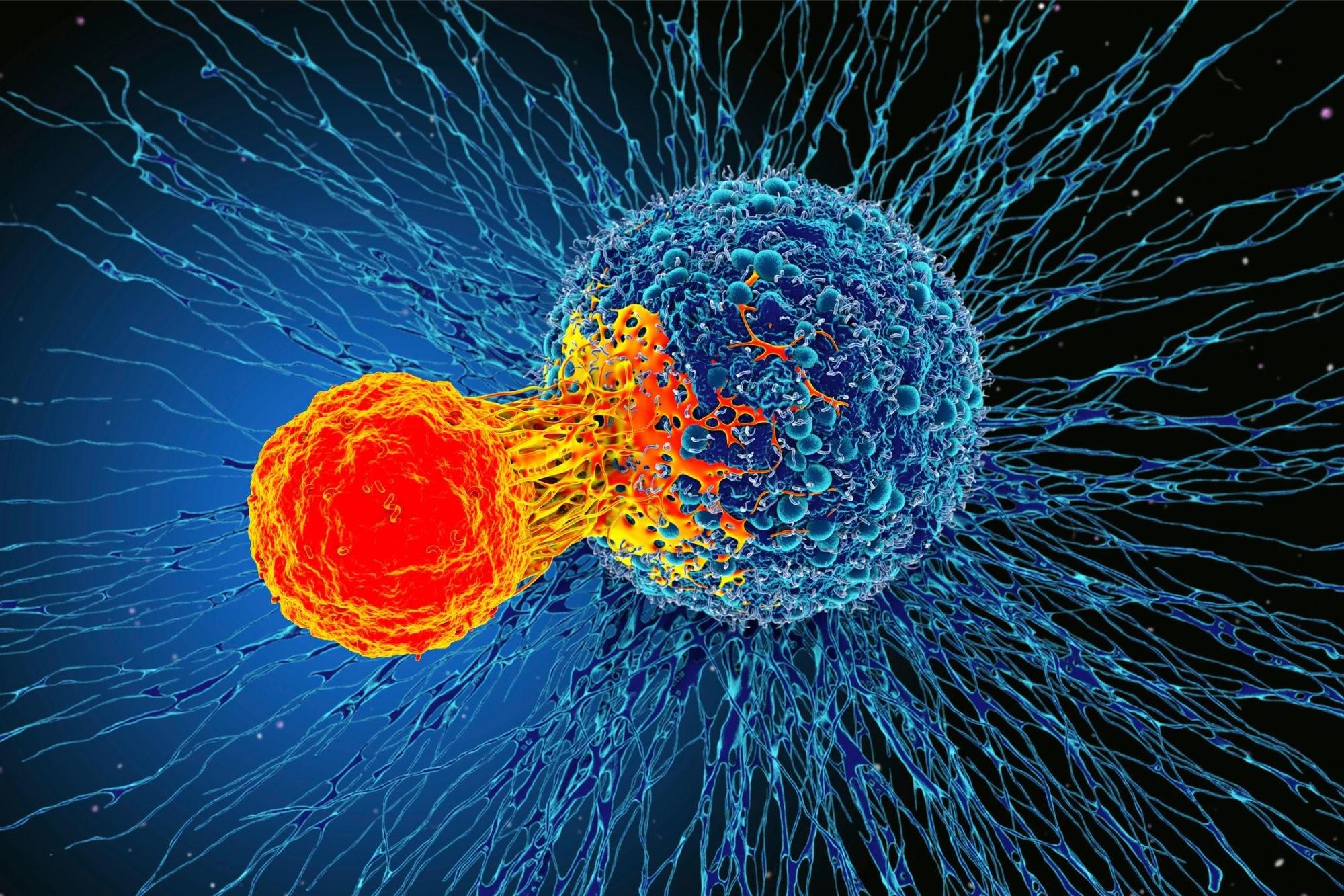
Toggle light / dark theme

Mapping the Thermal Forces That Push Particles through Liquids

Particles dispersed in a liquid typically jitter aimlessly in response to the random buffeting they receive from the molecules that surround them. But if the liquid is subjected to a steep temperature gradient, this random motion can become directional as the temperature gradient sets up flows that move the particles from hot regions of the liquid to colder ones. The theory of this so-called thermophoresis is actively developing, but direct observations of both the suspended particles and the liquid molecules are scant. Now Tetsuro Tsuji of Kyoto University in Japan and his colleagues have experimentally characterized the tiny surface flows that drive thermophoresis [1]. Those flows could be harnessed to move and concentrate DNA and other large biomolecules that are suspended in liquids.

For their experiments, the team glued a single polystyrene sphere, 7 µm in diameter, to the lid of a tiny transparent box. They filled the box with water laced with 500-nm-diameter fluorescent tracers. Shining a laser up through the bottom of the box, the team repeatedly drew a circle around the sphere, a process that trapped tracers located within the circle of light. The team focused a second laser, tuned to one of water’s absorption bands, at a spot 18 µm from the polystyrene sphere, locally heating the water to create a temperature gradient in the liquid and across the sphere.

Using a microscope the team observed that, after a few seconds, the tracers started flowing over the sphere’s surface, moving from the sphere’s cold end to its warmer one. From the observations, the researchers showed that this flow imparted momentum to the sphere. They also inferred the force that would have propelled the sphere away had it not been immobilized. Modeling the system under different conditions confirmed the inferences.

Tension Remodeling Resolves Tissue Architecture Question

A dynamical tension model captures how cells swap places with their neighbors in epithelial tissues, explaining observed phase transitions and cellular architectures.

Epithelial tissues line the surfaces of every organ in our bodies. In the earliest stages of organ development and in wound healing, the cells that make up these simple sheets constantly rearrange themselves, exchanging positions like molecules in a liquid. But this fluidization is often hindered by the formation of multicell clusters, whose origins remain unclear. Using a dynamical structural model, Fernanda Pérez-Verdugo and Shiladitya Banerjee of Carnegie Mellon University in Pennsylvania now identify the mechanical prerequisites that lead to the formation and dissolution of these stabilized clusters [1]. They show how dynamic feedback between tension and strain controls the tissue’s material properties.

Existing models of tissue fluidity treat epithelial tissues as foam-like, polygonal networks of cells whose edges join at triple points. However, these models fail to explain the mechanisms underpinning cell neighbor exchanges. In particular, they oversimplify such exchanges by treating them as an instantaneous process, thereby avoiding the impact of exchanges that stall midprocess. One resulting discrepancy with experimental results is the absence of stable “rosette” structures that are observed in developing tissues where four or more cells meet.

UK Biobank Releases Whole Genome Data from Half a Million People

After five years, more than 350,000 hours of genome sequencing, and over £200 million of investment, UK Biobank is releasing the world’s largest-by-far single set of human sequencing data—completing the most ambitious project of its kind ever undertaken. The new data, whole genome sequences of its half a million participants, will certainly drive the discovery of new diagnostics, treatments, and cures. Uniquely, the data are available to approved researchers worldwide, via a protected database containing only de-identified data.

This advance lies not only in the abundance of genomic data, but its use in combination with the existing data UK Biobank has collected over the past 15 years on lifestyle, whole body imaging scans, health information, and proteins found in the blood. The Pharma Proteomics Project was published last month in Nature, in the paper, “Plasma proteomic associations with genetics and health in the UK Biobank.

Looking forward, these data could be used to further advance efforts such as more targeted drug discovery and development, discovering thousands of disease-causing noncoding genetic variants, accelerating precision medicine, and understanding the biological underpinnings of disease.

Anthrobots: Scientists build tiny biological robots from human tracheal cells

Researchers at Tufts University and Harvard University’s Wyss Institute have created tiny biological robots that they call Anthrobots from human tracheal cells that can move across a surface and have been found to encourage the growth of neurons across a region of damage in a lab dish.

The multicellular robots, ranging in size from the width of a human hair to the point of a sharpened pencil, were made to self-assemble and shown to have a remarkable healing effect on other . The discovery is a starting point for the researchers’ vision to use patient-derived biobots as new therapeutic tools for regeneration, healing, and treatment of disease.

The work follows from earlier research in the laboratories of Michael Levin, Vannevar Bush Professor of Biology at Tufts University School of Arts & Sciences, and Josh Bongard at the University of Vermont in which they created multicellular biological robots from frog embryo cells called Xenobots, capable of navigating passageways, collecting material, recording information, healing themselves from injury, and even replicating for a few cycles on their own.

These Tiny, Wound-Healing Robots Start Life As Just 1 Human Cell

Regenerative medicine might just have had a new tool added to its arsenal: Scientists have created tiny biological robots out of living human cells. Though they may be small, the self-assembling bots are mighty, with a study demonstrating their potential for healing and treating disease.

The team had already proven their biological robotics chops back in 2020 with the creation of Xenobots, made from frog embryonic cells. They even managed to design Xenobots so that they could reproduce in a way that no living animal or plant does, something that had never been seen before.

The researchers weren’t sure whether the incredible capabilities of the Xenobots were in some way down to their amphibious origins, so they wanted to find out if biobots could also be created from the cells of other organisms. And why not begin with humans?

/* */