Colossal Biosciences claims three pups born recently are dire wolves, but they are actually grey wolves with genetic edits intended to make them resemble the lost species
Category: biotech/medical – Page 792
Your season of conception could influence how your body stores fat
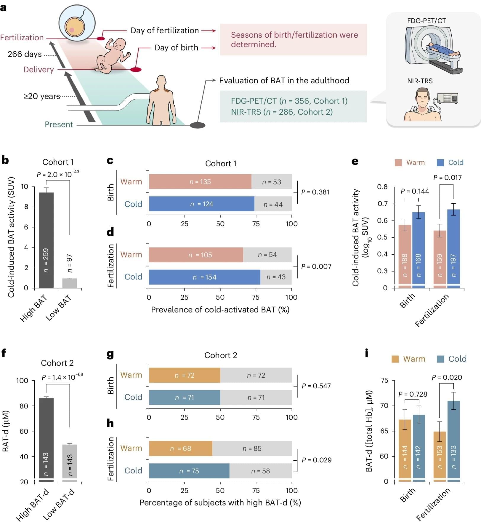
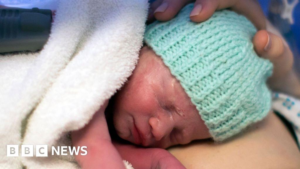
Individuals who were conceived in colder seasons are more likely to show higher brown adipose tissue activity, increased energy expenditure and a lower body mass index (BMI), and lower fat accumulation around internal organs, compared with those conceived in warmer seasons, suggests a study published in Nature Metabolism. The findings, based on an analysis involving more than 500 participants, indicate a potential role for meteorological conditions influencing human physiology.
Although eating habits and exercise are key indicators of fat loss, exposure to cold and warmth also plays a part. In colder temperatures, the body generates more heat (cold-induced thermogenesis) via brown adipose tissue activity and stores less fat in the form of white adipose tissue than it does in hotter temperatures. However, underlying factors contributing to individual differences in brown adipose tissue activity remain poorly understood, particularly in humans.
Takeshi Yoneshiro and colleagues analyzed brown adipose tissue density, activity and thermogenesis in 683 healthy male and female individuals between ages 3 and 78 in Japan, whose parents were exposed to cold temperatures (defined in the study as between 17 October and 15 April) or warm temperatures (between 16 April and 16 October) during the fertilization and birth periods.
Frustration incorporated: How mismatched geometries can enhance material strength and toughness
Anyone who’s ever tried tiling a floor, a backsplash or even an arts-and-crafts project probably knows the emotional frustration of working with pieces whose shapes don’t perfectly complement each other. It turns out, though, that some creatures may actually rely on similar mismatches to create geometric frustrations that result in complex natural structures with remarkable properties, such as protective shells and sturdy yet flexible bones.
Now, researchers at the University of Michigan have developed mathematical models showing one way that nature achieves this. These models, in turn, could help design advanced materials for medical devices, sustainable construction and more.
“Frustration—using these mismatched building blocks—gives rise to wonderful complexity and that complexity can be useful in providing superior material properties,” said Xiaoming Mao, U-M professor of physics and senior author of the new study.
Specialized Recycling System Eliminates Faulty Mitochondrial DNA
Damage to the mitochondria, the “power plants” of the cells, contributes to many diseases. Researchers from Heinrich Heine University Düsseldorf (HHU) and the University of Cologne led by HHU professor of medicine Dr David Pla-Martín, now describe in the scientific journal Science Advances how cells with defective mitochondria activate a special recycling system to eliminate damaged genetic material.
Damage to the genetic material of mitochondria – the mitochondrial DNA or mtDNA for short – can lead to diseases such as Parkinson’s, Alzheimer’s, amyotrophic lateral sclerosis (ALS), cardiovascular diseases and type 2 diabetes. Such damage also speeds up the ageing process. However, the cells are normally capable of identifying such damage and reacting.
How cells repair their mitochondria: Research uncovers a specialized recycling system
Damage to the genetic material of mitochondria—the mitochondrial DNA or mtDNA for short—can lead to diseases such as Parkinson’s, Alzheimer’s, amyotrophic lateral sclerosis (ALS), cardiovascular diseases and type 2 diabetes. Such damage also speeds up the aging process. However, the cells are normally capable of identifying such damage and reacting.
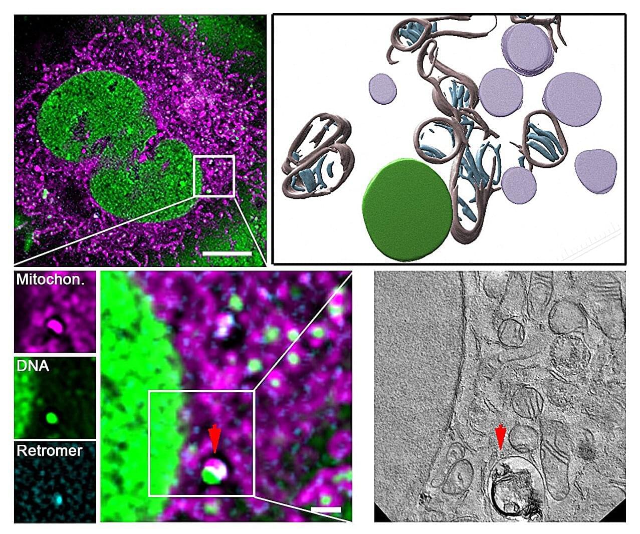
Scientists from University Hospital Düsseldorf and HHU have—in collaboration with the University of Cologne and the Center for Molecular Medicine Cologne (CMMC)—discovered a mechanism which protects and repairs the mitochondria. The research team, headed by Professor Pla-Martín from the Institute of Biochemistry and Molecular Biology I at HHU, has identified a specialized recycling system, which cells activate when they identify damage to the mtDNA.
According to the authors in Science Advances, this mechanism relies on a protein complex known as retromer and the lysosomes—cell organelles containing digestive enzymes. These special cellular compartments act like recycling centers, eliminating the damaged genetic material.
Gene discovery reveals potential for growing new heart arteries
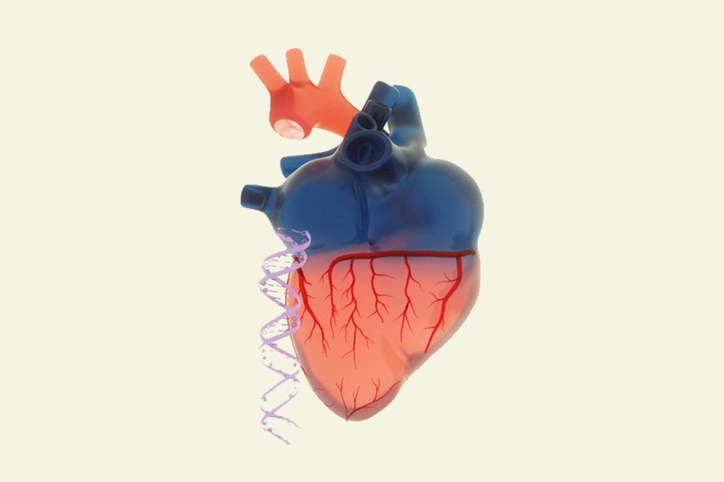
Most people have right-dominant hearts—which to a doctor or a researcher means they have an artery that extends from the right side of their hearts to supply oxygenated blood to the back side. For some people, this artery, called the posterior descending artery, comes from the left side or from both directions. A study has found that the gene CXCL12 is connected to this artery’s formation and that its directional pattern is set very early in human development.
The findings, reported in the journal Cell, represent a step toward developing “medical revascularization,” a long-term goal of Stanford researchers to create a treatment for blocked or limited-flow arteries by growing new ones to compensate.
“For the first time, we have evidence of a gene that regulates the development of one of the most important types of arteries in the human body,” said Kristy Red-Horse, co-senior author of the study and biology professor in the Stanford School of Humanities and Sciences. “And if we know the development pathways of these important arteries, then we can perhaps regrow them by reintroducing these pathways in a diseased heart.”
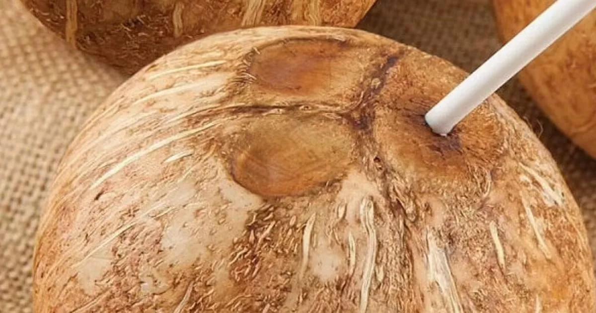
Man dies after sip from popular holiday drink left him with severe brain damage
Then, around three hours after he had drunk the coconut water, he started to have a fever with sweats and was vomiting. Paramedics came to his house and found him disorientated and pale while unable to balance.