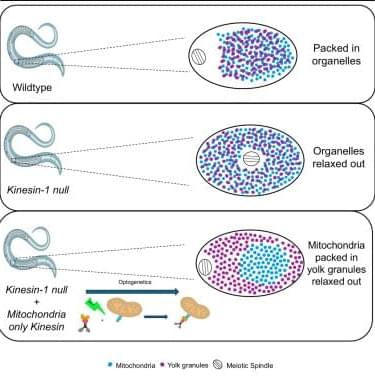
Aquino et al. find that kinesin-driven transport of mitochondria toward the center of a C. elegans oocyte is sufficient to drive movement of the meiotic spindle in the opposite direction to the periphery of the oocyte.
Category: biotech/medical – Page 782
A cloaked human stem-derived neural graft capable of functional integration and immune evasion in rodent models
Immune evasion of human stem-cell-derived neural graft in rodent models.
Transplantation rejection is the main challenge in human pluripotent stem cell (hPSC)-derived therapies.
The researchers used hPSC line (termed H1-FS-8IM), engineered to overexpress 8 immunomodulatory transgenes, to enable transplant immune evasion.
They show in co-cultures, H1-FS-8IM PSC-derived midbrain neurons evaded rejection by T lymphocytes, natural killer cells, macrophages, and dendritic cells.
The authors also provide preclinical evidence of pluripotent stem cell line evading immune detection after neural engraftment in a humanized immune system mouse model and reversal of motor symptoms in Parkinsonian rats.
Incorporation of a suicide gene within the universal donor cell ensures safety for cell-based therapies. https://sciencemission.com/A-cloaked-human-stem-cell-derived-neural-graft
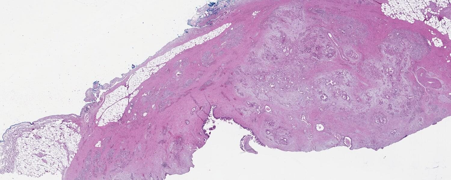
‘Sugar’ signatures help identify and classify pancreatic cancer cell subtypes
Van Andel Institute scientists and collaborators have developed a new method for identifying and classifying pancreatic cancer cell subtypes based on sugars found on the outside of cancer cells.
These sugars, called glycans, help cells recognize and communicate with each other. They also act as a cellular “signature,” with each subtype of pancreatic cancer cell possessing a different composition of glycans.
The new method, multiplexed glycan immunofluorescence, combines specialized software and imaging techniques to pinpoint the exact mix of pancreatic cancer cells that comprise tumors. In the future, this information may aid in earlier, more precise diagnosis.
Dr. Kilian Kelly, Ph.D. — CEO, Cynata Therapeutics — iPSC-Derived, Mesenchymal Stem Cell Therapies
Is Chief Executive Officer and Managing Director of Cynata Therapeutics (https://cynata.com/), a stem cell and regenerative medicine company that is known for its proprietary Cymerus platform, for the scalable and consistent production of mesenchymal stem cell (MSC)-based therapies.
Unlike traditional MSC therapies that rely on multiple donors, the Cymerus manufacturing process ensures that cells for therapeutic use can be produced in virtually limitless quantities from a single donor – making the opportunities endless and attractive from a manufacturing standpoint. The company has completed Phase I studies for Graft vs Host disease & Diabetic Foot Ulcers and have a number of Phase II, and even have a Phase III clinical trial, in progress.
Dr. Kelly has over 20 years’ experience in biopharmaceutical research and development, including almost 15 years focused on the development of mesenchymal stem cell (MSC) based therapies. He joined Cynata in March 2014, initially as Vice President, Product Development, then Chief Operating Officer from May 2019, and since July 2023 has been CEO & MD. At Cynata, he has overseen all stages of the development of the Cymerus induced pluripotent stem cell (iPSC)-derived MSC technology, including the first completed clinical trial of any iPSC-derived product worldwide.
Dr. Kelly previously held positions at Biota Pharmaceuticals, Mesoblast Limited, Kendle International, Amgen and AstraZeneca.
Dr. Kelly holds a Masters in Pharmacy degree from the Robert Gordon University, Aberdeen, a Ph.D. in Pharmaceutical Sciences from Strathclyde University, Glasgow, and he is a Graduate of the Australian Institute of Company Directors (AICD), Melbourne. He is a member of the International Society for Cell and Gene Therapy (ISCT), the International Society for Stem Cell Research (ISSCR), the Royal Pharmaceutical Society and the AICD.
Dr. Kelly also serves on the ISCT Asia-Pacific Industry Committee, the ISSCR Best Practices Working Group for the Development of PSC-Derived Therapies and the Industry Interface Committee of the Center for Commercialization of Regenerative Medicine (CCRM) Australia.
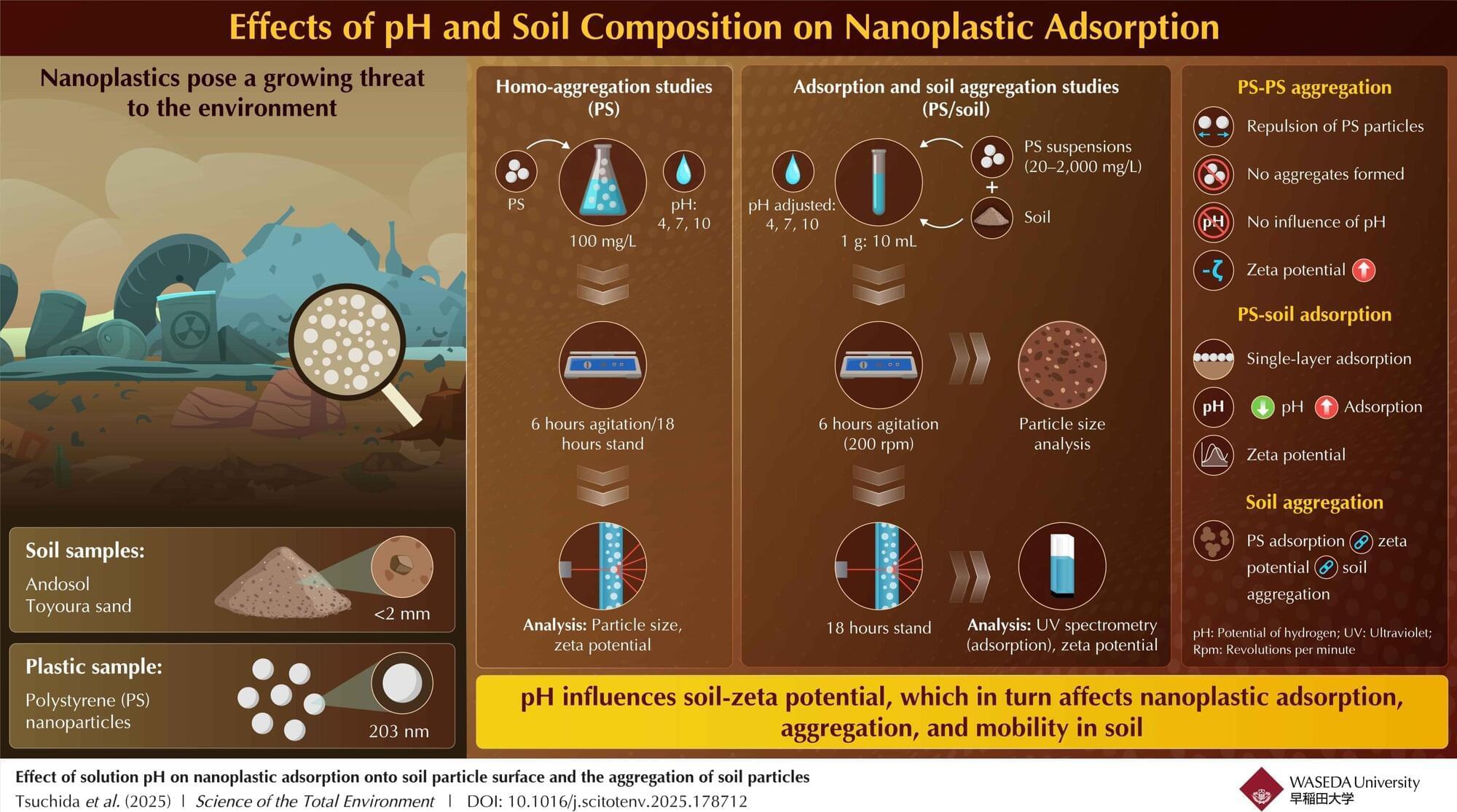
Nanoplastics in soil: How soil type and pH influence mobility
Plastics are everywhere—from packaging and textiles to electronics and medical devices. As plastic waste breaks down, it releases microscopic particles that can penetrate our ecosystems, hinder plant growth, and potentially transfer harmful pollutants to organisms, including humans. Therefore, these plastic particles are a potential threat to the ecosystem, especially in their nanoparticulate form (1–100 nm diameter), which can penetrate the environment through different routes, including the soil beneath our feet.
With this in mind, a team of researchers from Japan set out to study the migration behavior of nanoplastics in different soil types. The study was led by Kyouhei Tsuchida, a Ph.D. student from the National Institute of Advanced Industrial Science and Technology (AIST) and Waseda University, Japan, with fellow students Yukari Imoto, Takeshi Saito, and Junko Hara also from AIST, and Professor Yoshishige Kawabe from the Department of Resources and Environmental Engineering, Waseda University. This study was published online in the journal Science of the Total Environment on April 4, 2025.
The researchers focused on the adsorption of the nanoplastics on soil and the aggregation characteristics of both the nanoplastics and soil particles under varying pH conditions. “The aggregation properties of nanoplastics and their adsorption onto soil particle surfaces are known to affect their migration in soil,” notes Tsuchida. “We conducted experiments to analyze these traits to get a better understanding of the migration of nanoplastics.”
Japanese scientists use stem cell treatment to restore movement in spinal injury patients
A stem cell treatment helped improve the motor function of two out of four patients with a spinal cord injury in the first clinical study of its kind, Japanese scientists said.
There is currently no effective treatment for paralysis caused by serious spinal cord injuries, which affect more than 150,000 patients in Japan alone, with 5,000 new cases each year.
Researchers at Tokyo’s Keio University are conducting their study using induced pluripotent stem cells (iPS)—created by stimulating mature, already specialized, cells back into a juvenile state.

Japan team carries out world-first spinal cord stem cell trial
A Japanese university said Friday it has successfully transplanted stem cells into a patient with a spinal cord injury, in the first clinical trial of its kind.
There is currently no effective treatment for paralysis caused by serious spinal cord injuries, believed to affect more than 100,000 people in Japan alone.
Surgeons at Tokyo’s Keio University want to study whether induced pluripotent stem (iPS) cells can be used to treat the injuries.
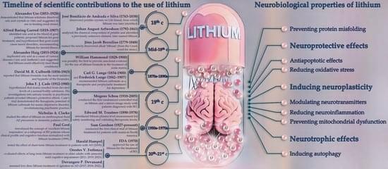
An Overview of the Effects of Lithium on Alzheimer’s Disease: A Historical Perspective
Lithium was introduced into psychiatric practice in the late nineteenth century and has since become a standard treatment for severe psychiatric disorders, particularly those characterized by psychotic agitation. It remains the most effective agent for managing acute mania and preventing relapses in bipolar disorder. Despite potential adverse effects, lithium’s use should be carefully considered relative to other treatment options, as these alternatives may present distinct safety and tolerability profiles. The World Health Organization classifies lithium salts as ‘essential’ medications for inclusion in global healthcare systems. Over the past two decades, the growing recognition of lithium’s efficacy—extending beyond mood stabilization to include reducing suicide risk and inducing neuroprotection—has led to its incorporation into clinical practice guidelines.